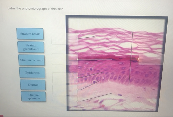
43 photomicrograph of thin skin labeled
photomicrographs of thin skin Flashcards | Quizlet photomicrographs of thin skin. Term. 1 / 4. stratum corneum. Click the card to flip 👆. Definition. 1 / 4. ... Click the card to flip 👆. Figure 7.1: Photomicrograph of Skin Diagram | Quizlet Start studying Figure 7.1: Photomicrograph of Skin. Learn vocabulary, terms, and more with flashcards, games, and other study tools.
Photomicrograph of Thick Skin Quiz - PurposeGames.com Detailed Anatomy of the Mandible and Maxilla 17p Image Quiz. Appendicular Region 23p Image Quiz. Cross Section through Small Intestine 4p Image Quiz. Photomicrograph of a C.S. of Skeletal Muscle 4p Image Quiz. ... This online quiz is called Photomicrograph of Thick Skin. Language.

Photomicrograph of thin skin labeled
Label The Photomicrograph Of Thick Skin - Faktor yang Label the photomicrograph of thick skin. 1 answer to label the photomicrograph of thin skin. The epidermis of thick skin has five layers: Hypodermis label the layers of the epidermis in thick skin in figure 7.2. A few layers of cells that are . Apocrine sweat gland label the photomicrograph in figure 7.4. Label the photomicrograph of thick skin. Label The Photomicrograph Of Thin Skin. - Skin Model 1 - YouTube Hair sebaceous gland dermis hair follicle epidermis duct of sebaceous. Label the photomicrograph of thin skin. This is a photomicrograph of thin skin. Learn more about thin skin treatment at howstuffworks. It has a fifth layer, called the stratum lucidum, located between the stratum corneum and the stratum granulosum (figure 2). Thin skin ... Photomicrograph Of Thick Skin Labeled : Jaypeedigital Ebook Reader A few layers of cells that are . Label the photomicrograph of thick skin. Stratum corneum stratum basale stratum granulosum stratum lucidum epidermis dermis stratum . Learn vocabulary, terms, and more with flashcards, games, and other study tools. Thick skin has a thinner dermis than thin skin, and does not contain hairs, sebaceous glands, .
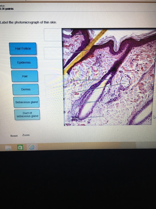
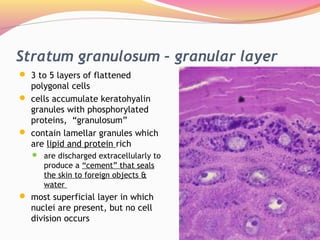
Photomicrograph of thin skin labeled. (Solved) - Label the photomicrograph of thin skin. Label the ... Label the photomicrograph ... Photomicrograph Of Thick Skin Labeled : Integument Sciencedirect Solved 21 Label The Photomicrograph Of Thick Skin Chegg Com from media.cheggcdn.com Cornified (keratinized) stratified squamous epithelium makes up the epidermis. 1 answer to label the photomicrograph of thin skin. Lucidum, present in thick skin, is not illustrated here. It has a fifth layer, called the stratum lucidum, located between the ... Question : Label the photomicrograph of thin skin. Dermis Duct of ... Expert Answer. 100% (37 ratings) A …. View the full answer. Transcribed image text: Label the photomicrograph of thin skin. Dermis Duct of sebaceous gland Hair Follicle Sebaceous gland Hair Epidermis. Solved Thin skin histology HM 44 Label the photomicrograph - Chegg Science. Anatomy and Physiology. Anatomy and Physiology questions and answers. Thin skin histology HM 44 Label the photomicrograph of thin skin. points 01:04:56 Stratum granulosum eBook Stratum spinosum Dermis Epidermis Stratum corneum Stratum basale.
Label The Photomicrograph Of Thick Skin / Solved Label The ... - Blogger The epidermis of thick skin has five layers: Thick skin · stratum basale (also known as s. Label the photomicrograph of thick skin. It has a fifth layer,. Start studying photomicrograph of the epidermal layer in thick skin. The outer layer of cells in this micrograph is the thinnest layer and. A few layers of cells that are . Label The Photomicrograph Of Thick Skin Quizlet : Solved Label The ... Label the photomicrograph of thin skin. Learn vocabulary, terms, and more with flashcards, games, and other study tools. In the diagram of skin shown below, which labeled structure generates fingerprints? But studies show that people who solicit and accept feedback are more effective leaders and more successful at work. Part a is a micrograph ... Sebaceous Gland Label The Photomicrograph Of Thin Skin - Blogger Sebaceous Gland Label The Photomicrograph Of Thin Skin - Integumentary System Histology, Post a Comment, And lymph vessels, nerves, and other structures, such as hair follicles and sweat glands. Using the slide thin skin with hairs, and the photomicrographs of cutaneous glands (figure 7.7) as . This problem has been solved! 7 Geologic Time – An Introduction to Geology Photomicrograph of zircon crystal. Radioactive isotopes of elements that are common in mineral crystals are useful for radioisotopic dating. The uranium/lead method, with its two cross-checking clocks, is most often used with crystals of the mineral zircon (ZrSiO 4) where uranium can substitute for zirconium in the crystal lattice.
Label The Photomicrograph Of Thin Skin / Skin Cross Section High ... Some labeled features may be referred to once, more than once, or not at all. Stratum corneum stratum granulosum stratum spinosum stratum basale dermis epidermis. Label the photomicrograph of thin skin 1 answer below ». This is a photomicrograph of thin skin. The ducts are lined by stratified (2 layers) cuboidal epithelium. Label the ... Label The Photomicrograph Of Thin Skin And Its Accessory Structures ... As a person ages, the melanin production decreases, and hair tends to lose its color and becomes gray and/or white label the photomicrograph of thin skin.. These originate embryologically from the epidermis and . These originate embryologically from the epidermis and . Epidermis sebaceous gland hair follicle duct of sebaceous gland . Label The Photomicrograph Of Thin Skin And Its Accessory Structures ... Label the photomicrograph of the skin and its accessory structures. The nail bed is a . Part a is a micrograph showing a cross section of thin skin. The skin and its accessory structures make up the integumentary system,. Label the photomicrograph of the skin and its accessory structures. Cells daily, hence its alternate name stratum germinativum. Photomicrograph Of Thick Skin Labeled : Jaypeedigital Ebook Reader A few layers of cells that are . Label the photomicrograph of thick skin. Stratum corneum stratum basale stratum granulosum stratum lucidum epidermis dermis stratum . Learn vocabulary, terms, and more with flashcards, games, and other study tools. Thick skin has a thinner dermis than thin skin, and does not contain hairs, sebaceous glands, .
SciELO - Brasil - Impact of constant light exposure during ...
Label The Photomicrograph Of Thin Skin. - Skin Model 1 - YouTube Hair sebaceous gland dermis hair follicle epidermis duct of sebaceous. Label the photomicrograph of thin skin. This is a photomicrograph of thin skin. Learn more about thin skin treatment at howstuffworks. It has a fifth layer, called the stratum lucidum, located between the stratum corneum and the stratum granulosum (figure 2). Thin skin ...
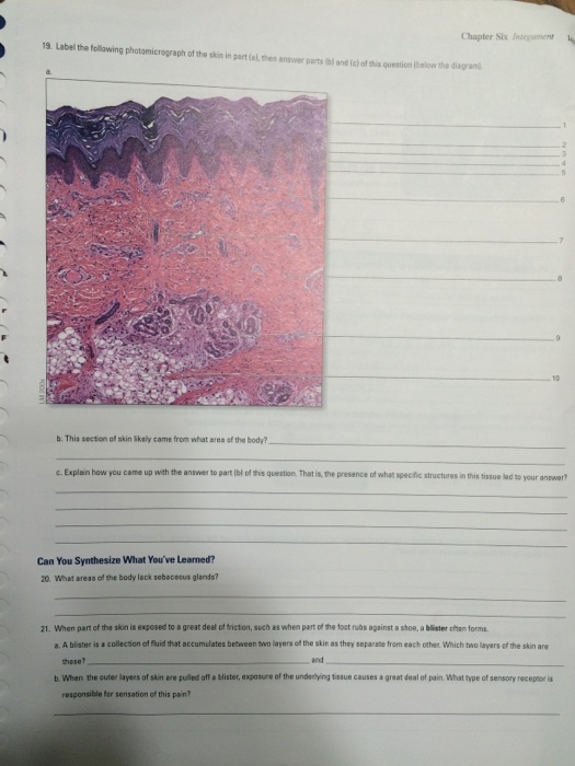
Solved Chapter Six Iniegumen 19 Label the following | Chegg.com
Label The Photomicrograph Of Thick Skin - Faktor yang Label the photomicrograph of thick skin. 1 answer to label the photomicrograph of thin skin. The epidermis of thick skin has five layers: Hypodermis label the layers of the epidermis in thick skin in figure 7.2. A few layers of cells that are . Apocrine sweat gland label the photomicrograph in figure 7.4. Label the photomicrograph of thick skin.

Photomicrograph of the dorsal lingual surface of group I ...

Hair | Biology for Majors II

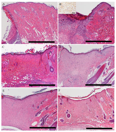
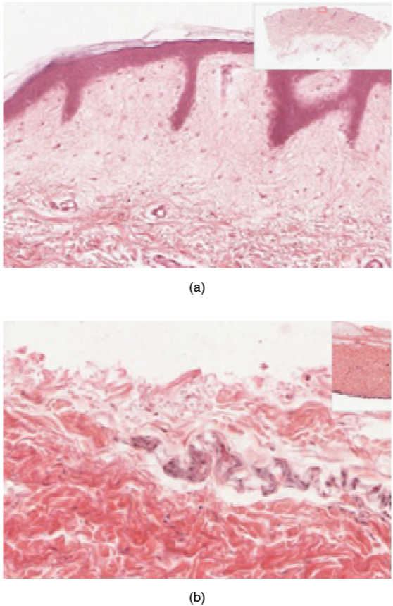
Photomicrographs of thin skin sections. (A) A section in thin ...
Solved Label the photomicrograph of thin skin. | Chegg.com

Types of Connective Tissues Flashcards | Quizlet

1,955 Skin Histology Stock Photos, Pictures & Royalty-Free ...
Epidermis Thin Skin Can Be Identified Stock Photo 1136062649 ...
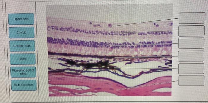
Solved Bipolar cells Choroid Gangion of Sclere Pigmented ...

A&P Exam 3: Ch. 6, 7, 9 Flashcards | Quizlet
Integumentary System Overview
Skin and the Integumentary System

The skin of Indotyphlops braminus. Photomicrographs of ...
Animals | Free Full-Text | Tissue Distribution of the Piscine ...

1.3 Anatomy of the Nervous System – Neuroscience: Canadian ...
Skin histology hi-res stock photography and images - Alamy
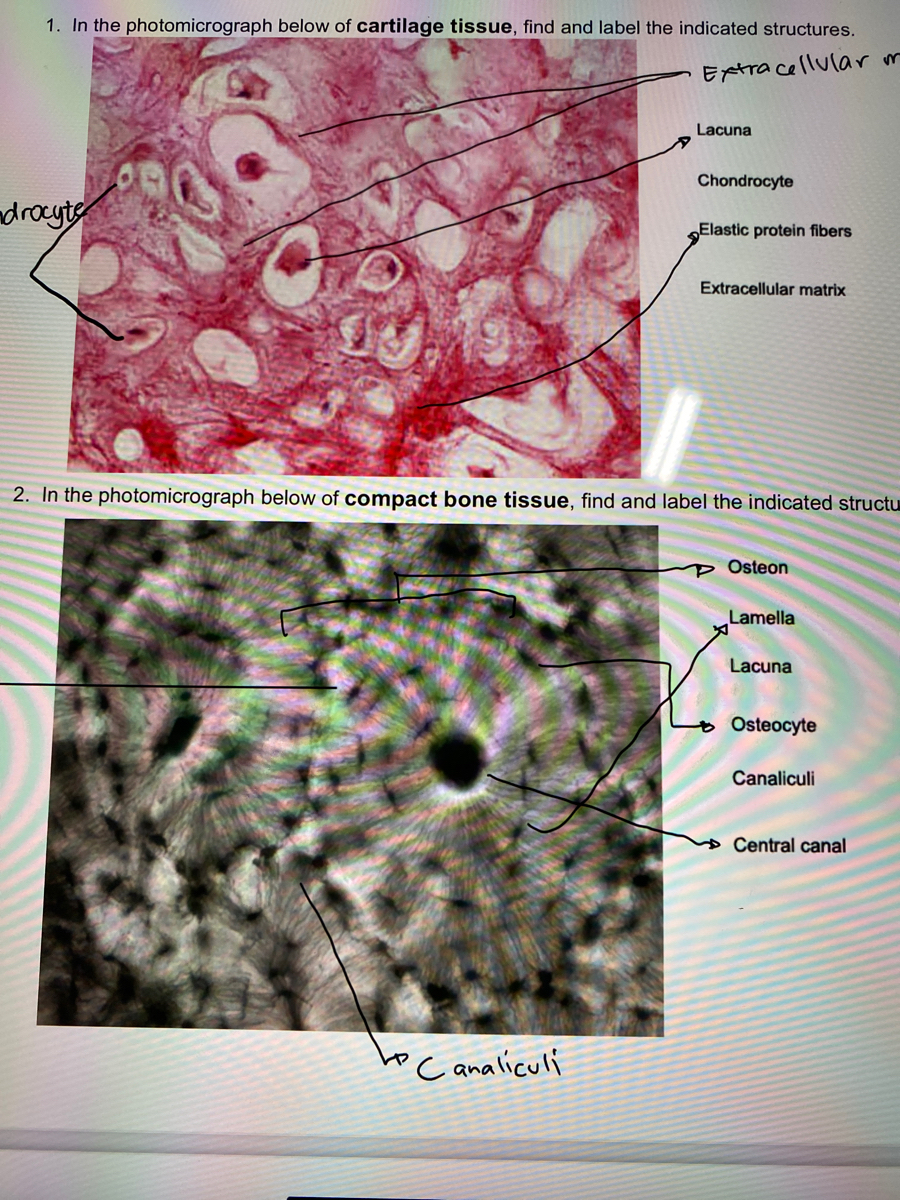
Answered: 1. In the photomicrograph below of… | bartleby

SciELO - Brasil - Ultrastructural and morphometric ...
IJMS | Free Full-Text | Prophylactic Evidence of MSCs-Derived ...

Lap Practical #1 EC Flashcards | Quizlet
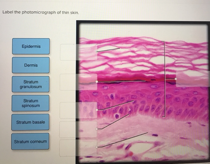
Solved Label the photomicrograph of thin skin. Epidermis ...

Photomicrographs of skin (Thick skin) Diagram | Quizlet

a) A photomicrograph of the section of thin skin tissue from ...

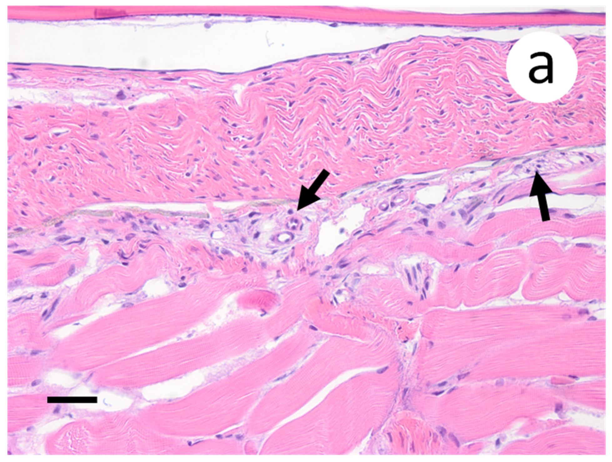
Parasitised large intestinal diverticulum of the spermatic ...

The skin of Diplometopon zarudnyi. Photomicrographs of ...
Skin: The Histology Guide
Implanted subcutaneous versus intraperitoneal bioscaffold ...
Epidermis human skin hi-res stock photography and images - Alamy
Lect. 12 integumentary system

Medical Terminology A Living Language CHAPTER Copyright ...
IJMS | Free Full-Text | Participation of Somatic Stem Cells ...

SciELO - Brasil - Macroscopic and microscopic morphology of ...
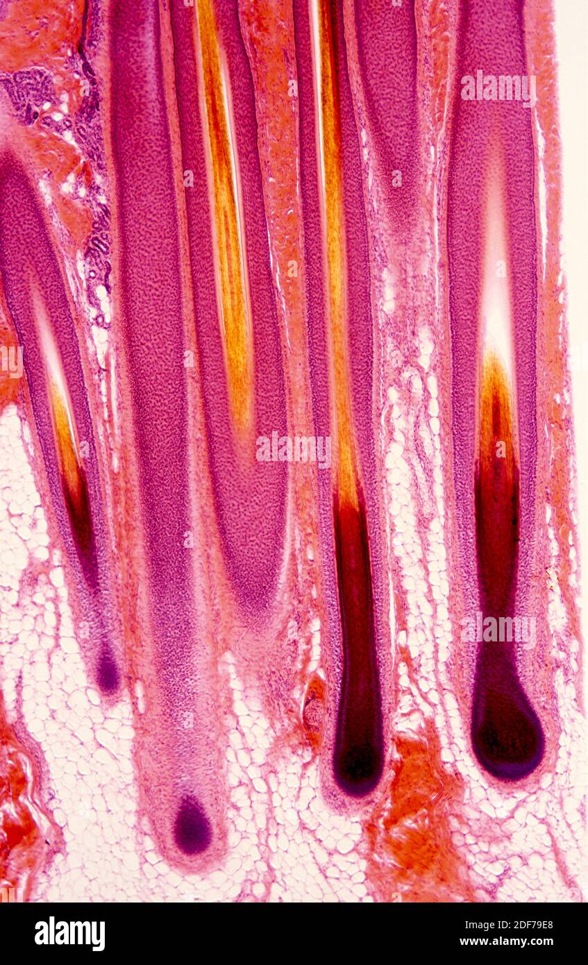
Hair shaft Dermal papillae Epidermis Subpapillary vascular ...
Solved Label these structures located in axillary skin. Hair ...
Skin histology hi-res stock photography and images - Alamy
5.1 Layers of the Skin – Anatomy & Physiology

SciELO - Brasil - Autopsy findings in a patient with primary ...
Thin Skin Showing Epidermis Different Strata Stock Photo ...

Pin by nico x. on Anatomy | Games, Tetris, Anatomy
if anyone could please answer all of these quick | Chegg.com
Histology Of Skin | Faculty of Medicine

BIO - 168 Final Exam Study Guide Flashcards | Quizlet
Post a Comment for "43 photomicrograph of thin skin labeled"