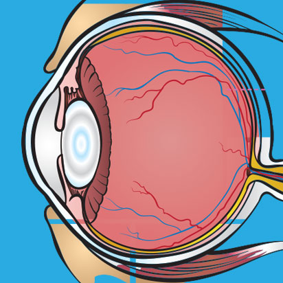
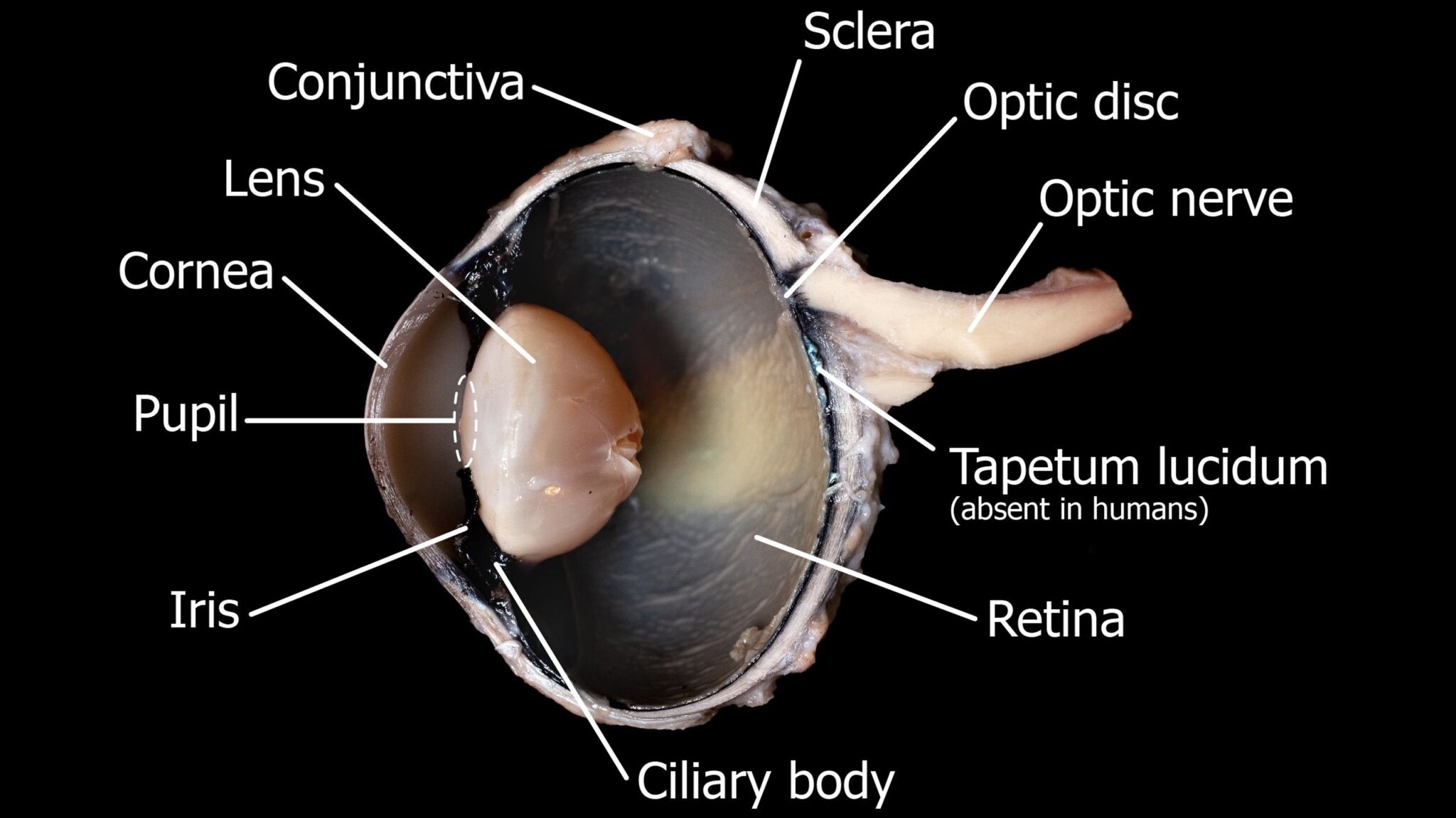
45 labeled cow eye dissection
Cow Eye Dissection Lab from Anatomy and Physiology Cow's Eye Dissection Lab Page 2. List the two functions of the lens. The lens is a transparent structure in the eye that, along with the cornea, helps to refract and focus light. objects further away. Describe the iris and explain its function. The iris is the colorful part of the eye. Gastrointestinal tract - Wikipedia The lower gastrointestinal tract includes most of the small intestine and all of the large intestine. In human anatomy, the intestine (bowel, or gut.Greek: éntera) is the segment of the gastrointestinal tract extending from the pyloric sphincter of the stomach to the anus and as in other mammals, consists of two segments: the small intestine and the large intestine.
University of South Carolina on Instagram: “Do you know a future ... Web13.10.2020 · Do you know a future Gamecock thinking about #GoingGarnet? 🎉 ••• Tag them to make sure they apply by Oct. 15 and have a completed application file by Nov. 2 to get an answer from @uofscadmissions by mid-December. 👀 // #UofSC

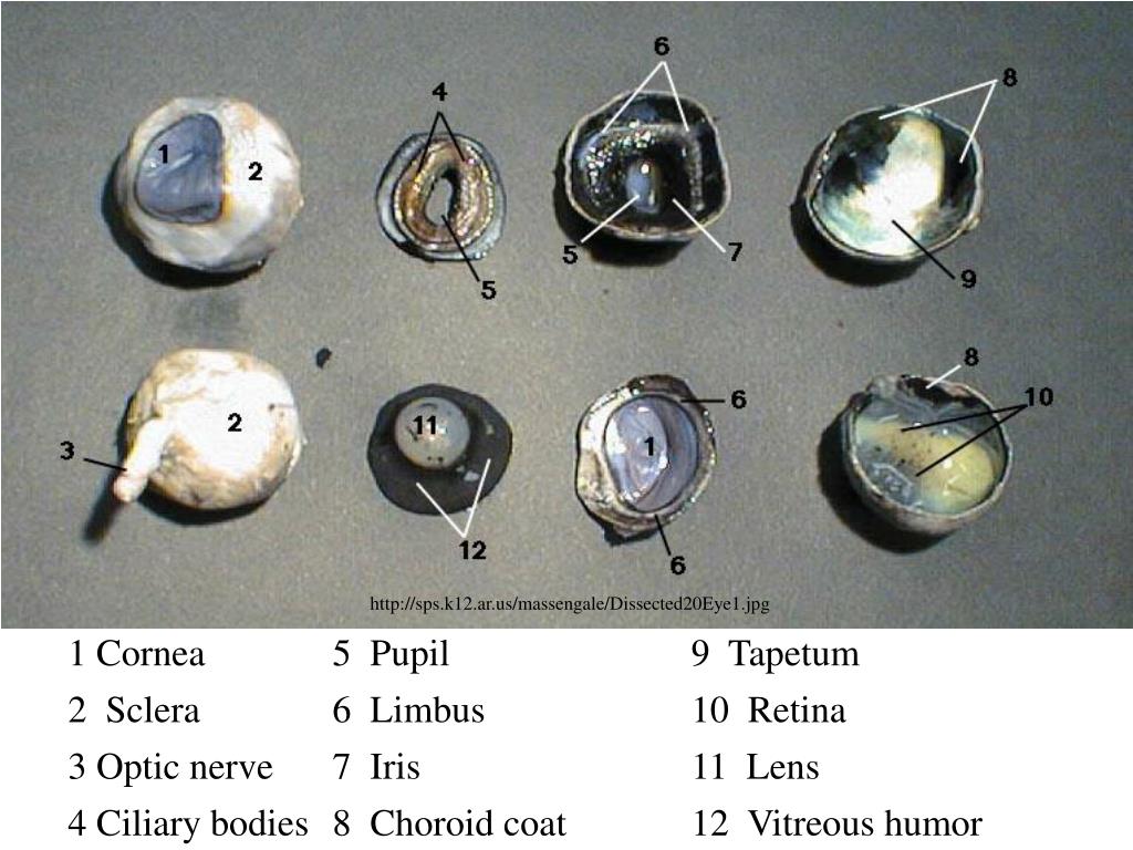
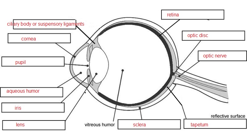
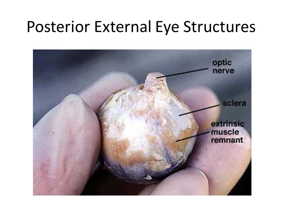
Labeled cow eye dissection
Cow Eye Dissection Flashcards | Quizlet A ring of muscle tissue that forms the colored portion of the eye around the pupil and controls the size of the pupil opening. Lens The transparent structure behind the pupil that changes shape to help focus images on the retina. Retina Located in the back of the eye, contains the rods and cones. pupil Dissecting An Eyeball - Krieger Science Returning to our dissection, underneath the retina is a pretty, shiny, blue-green mirror, officially called the tapetum. This is what causes cow's eyes to shine in headlights, and it is probably the most memorable part of an eye dissection for children. This is also one thing that cows and people do not have in common. (PDF) LEWIN'S GENES XII | Mtro. Raúl Reyes Bautista WebHow did evidence from the study of metabolic defects contribute to the fundamental relationship between genes and proteins? Garrod first suggested that genes dictate phenotypes through enzymes that catalyze specific chemical reactions.
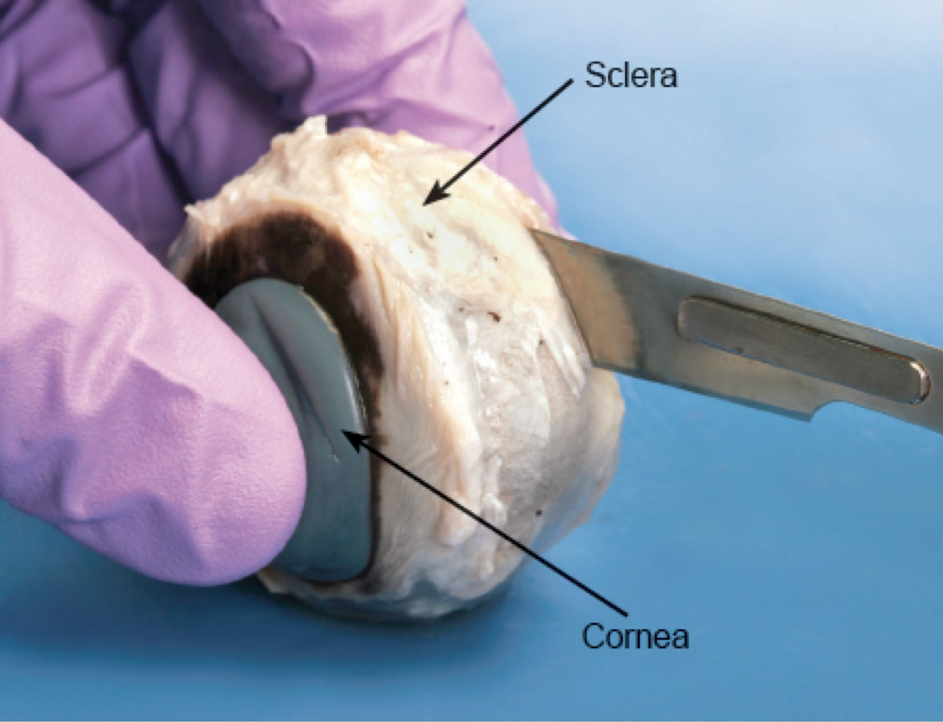
Labeled cow eye dissection. Cow Eye Dissection Guide - Google Slides Cow Eye. Use the point of a scissors or a scalpel to make an incision through the layers of the eye capsule (similar to figure 1); there are three layers from the exterior: sclera, whitish/grey, continuous with the transparent cornea, choroid, thin dark black layer and the retina, thin greyish/pink layer. Use a scissors to dissect the entire ... Read Book Anatomy Lab Cow Heart Dissection Answer Key 1 Sheep Brain and Cow Eye Dissection Lab Report Ivy Tech Anato-my and Physiology 101 2/27/2020 Abstract The purpose of the sheep brain and cow eye dissection is to familiarize locating and identify the regions and structures in the brain and eye. The sheep brain and cow eye were used because their functions are similar of a human brain and eye. Cow's Eye Dissection - Eye diagram - Exploratorium A cow's pupil is oval. A tough, clear covering over the iris and the pupil that helps protect the eye. Light bends as it passes through the cornea. This is the first step in making an image on the retina. The cornea begins bending light to make an image; the lens finishes the job. The layer of light-sensitive cells at the back of the eye. Cow Eye Dissection & Anatomy Project | HST Learning Center Cow Eye Dissection: Internal Anatomy 1. Place the cow's eye on a dissecting tray. The eye most likely has a thick covering of fat and muscle tissue. Carefully cut away the fat and the muscle. As you get closer to the actual eyeball, you may notice muscles that are attached directly to the sclera and along the optic nerve.
Well - The New York Times WebBringing Home Some ‘Hairy Joie de Vivre,’ and Taking Notes. Like many, the canine behavioral expert Alexandra Horowitz adopted a dog during the pandemic. Cow eye - dissection and label Cow eye shown with labeled cornea. The cornea is the transparent front part of the eye that covers the iris, pupil, and anterior chamber. The cornea, with the anterior chamber and lens, refracts light, with the cornea accounting for approximately two-thirds of the eye's total optical power. 3. Home Page: The Journal of Pediatrics Web04.12.2019 · The Journal of Pediatrics is an international peer-reviewed journal that advances pediatric research and serves as a practical guide for pediatricians who manage health and diagnose and treat disorders in infants, children, and adolescents.The Journal publishes original work based on standards of excellence and expert review. The Journal … Amazon.com: Cow Eyes For Dissection 470221-070 - 1.5 to 3" Pure Preserved Cow Eyes for Classroom Dissection - Pack of 1 Preserved Cow Eye 4.5 out of 5 stars2 $6.15$6.15 Get it Tue, Oct 4- Fri, Oct 7 FREE Shipping Perfect Solution Preserved Cow Eye, Plain, Bulk Bag 4.6 out of 5 stars20 470158-798 - Formalin Preserved Cow Eyes - Bovine Eyes - Each 5.0 out of 5 stars1 $48.67$48.67
Lab 10—Labeled Cow Eye The Cow Eye (Labeled) Return to: Lab 10 Page BIO 137 Main Page Be sure to practice using the unlabeled images. Coronal section, anterior view (lens and vitreous humor displaced) Sagittal section: This page created and maintained by Udo M. Savalli. Last updated April 18, 2006. Cow Eye dissection Free Essay Example - StudyMoose The purpose of this lab was to explore the anatomy of a cow eye. The eye is a special sensory organ with a complex structure and function. The objective was to gain a better understanding of the structure and the function of the eye. In this lab, we set out to dissect a cow's eye and identify the different parts of the eye. Hypothesis (PDF) Nanda NIC NOC | dwi adiyanto - Academia.edu WebAcademia.edu is a platform for academics to share research papers. Cow Eye Dissection PowerPoint Presentation, free download - SlideServe Cow Eye Dissection. Objectives By the end of this lesson, students will be able to: • Demonstrate proper dissection safety techniques. • Explore and identify the external anatomy of the cow eye. • Explore and identify the internal anatomy of the cow eye. • Compare and contrast human and cow eye anatomy • Evaluate how the structure of ...
PDF Cow Eye Dissection Guide - cbsd.org DISSECTION OF THE COW EYE Please make sure to wear gloves and safety glasses when you are dissecting, and make sure to clean up thoroughly after the lab. Also, the cow eyes can be rather slippery, so use caution when handling and cutting them. You will need a scalpel and forceps. 1. First, identify the most external structures of the eye.
Cow Eye Dissection - The Biology Corner COW EYE DISSECTION 1. Examine the outside of the eye. You should be able to find the sclera, or the whites of the eye. This tough, outer covering of the eyeball has fat and muscle attached to it 2. Locate the covering over the front of the eye, the cornea. When the cow was alive, the cornea was clear.
Video News - CNN WebWatch breaking news videos, viral videos and original video clips on CNN.com.
Cow Eye Dissection Pre-lab Key - Google Docs 1. Use the structures listed in question #2 and label the diagram. 2. Describe the function of the following structures: Cornea - tough covering over the iris that helps protect the eye and directs the light towards the lens. Optic nerve- The bundle of nerve fibers that carries the impulse to the brain.
Cow Eye Dissection - YouTube About Press Copyright Contact us Creators Advertise Developers Terms Privacy Policy & Safety How YouTube works Test new features Press Copyright Contact us Creators ...
Cow Eye Dissection Kit for Kids Animal Anatomy Labs | HST Cow Eye Dissection Kit $9.95 This Cow Eye Dissection Kit gives an inside view of how the eye works. It comes with everything you need for this activity, including a preserved cow eye specimen, a step-by-step dissection guide & essential dissection tools. quantity Ages 11+ In Stock & Ready to Ship Need it fast? See delivery options in cart.
PDF Cow Eye Dissection - University of Illinois Urbana-Champaign Cow Eye Dissection May 2013 2 Dissection equipment should be washed, and work spaces should be wiped down with paper towels and a spray cleaner. Once cleaned spaces are approved by the facilitator, students should dispose of gloves, clean and replace their goggles, and immediately wash their hands at the nearest sink. IV.
Cow Eye Dissection & Parts of the Eye Diagram | Quizlet Photoreceptor cells in the eye that detect black, white, and gray cones Photoreceptor cells in the eye that detect color aqueous humor Fluid in front chamber of eye; nourishes the cornea and the lens. vitreous humor The clear gel filling the space of the eyeball between the lens and the retina Sets with similar terms The eye LadyBuchanan1
anatomy and physiology of cow - Microsoft anatomy sheep dissection heart side dissected pig physiology science human class card labeled medical septum cow notes parts interventricular study. Pin On Anatomy And Physiology . eye worksheet diagram human cow dissection anatomy worksheets structure physiology answers eyeball drawing label body labeling eyes ear template muscle
PDF Cow Eye Dissection: Examining Structure and Function - Woodstown During this activity, you will dissect a cow eye. You will observe several important features of the eye and develop your understanding of how each part functions to make vision possible. Materials • Preserved Cow Eye • Scalpel or Scissors • Forceps • Dissection Tray • Gloves • Safety Glasses • Lab Apron 1.
NFL Football Highlights, Clips & Analysis | NFL.com WebThe destination for all NFL-related videos. Watch game, team & player highlights, Fantasy football videos, NFL event coverage & more
Dissection of Cow eye | Course Hero ANATOMY ANT. Dissection of Cow eye. Dissection of Cow eye. School Broward College; Course Title ANATOMY ANT; Type. Lab Report. Uploaded By Marchee3Bass. Pages 1 This preview shows page 1 out of 1 page. View full document ...
Oxytetracycline | C22H24N2O9 - PubChem Oxytetracycline is a tetracycline used for treatment of infections caused by a variety of Gram positive and Gram negative microorganisms including Mycoplasma pneumoniae, Pasteurella pestis, Escherichia coli, Haemophilus influenzae (respiratory infections), and Diplococcus pneumoniae.
[Solved] How to label a cow eye dissection? | Course Hero When labeling a cow eye dissection, it is also important to use clear and concise labels. Use abbreviations if necessary, but make sure that the labels are still understandable. For example, you might label the cornea as "C" and the iris as "I". In general, it is best to label the parts of the eye before you start the dissection.
[カテゴリ未分類]の記事一覧 | わたしのブログ by Dmdilpo -... xamii lvcwtlpmn. Galore tube What is a good gamebattles logo maker Cats made out of keyboard symbols Stephine abrams Simple piano notes for poker face Sterlings embroidery san antonio west ave Mario scene builder Virtual human dissection games Naughty dares to ask a guy over text Create ecomap online for mac Floor candle stands Sadlier-oxford vocabulary workshop cheats Virtual c-section games ...
Cow Eye Dissection Teaching Resources | Teachers Pay Teachers Cow Eye Dissection by CrazyScienceLady 49 $5.00 PDF Cow Eye Dissection: Directions and questions for dissecting a cow eye. Also includes a diagram of the eye, fun facts, an activity on how to find your blind spot and all answer keys. Appropriate for grades 4-7, I run this lab for each of the 4th grade classes at my school. It's a HUGE hit!!!
Cow Eye Dissection | Carolina.com We offer a full range of dissecting equipment to fit all your lab needs. There are sets available for all skill levels or can be customized. Lab Equipment Carolina is your quality source for a well-equipped lab. Take time to view our high quality science lab equipment that has proven durability to handle any lab activity. Life Science Life Science
PDF Cow Eye Dissection Lab - Home Science Tools This cow eye dissection kit comes with everything you need to conduct a lab examination. Safety Guidelines • Work in a place separate from eating and food preparation areas. • Use disposable latex gloves or nitrile gloves during the dissection and cleanup. • Use only dissection tools provided.
PDF COW'S EYE dissection - Exploratorium COW'S EYE dissection page 6 Now take a look at the rest of the eye. If the vitreous humor is still in the eyeball, empty it out. On the inside of the back half of the eyeball, you can see some blood vessels that are part of a thin fleshy film. That film is the retina. Before you cut the eye open, the vitreous humor
Cow Eye and Sheep Brain Dissection Report by Jaymee Brockhaus Sheep is an animal whose brain is noticeably similar to the human brain. One difference is that the sheep does not have the ability to think, and that simply makes their brain less efficient than the human brain. By dissecting and examining the anatomy of a preserved cow eye, you can learn how your own eye forms images and sends these images to ...
Wellness Library | Cigna WebEye Problems: Using Eyedrops and Eye Ointment. Eyes and Vision. F FA. Face-Lift (Rhytidectomy) Facial Injuries . Facial Problems, Noninjury. Failure to Thrive. Fainting. Fallopian Tube Procedures for Infertility. Family Life Cycle. Family Therapy. Farsightedness (Hyperopia) Fasting. Fatigue (PDQ®): Supportive care - Health Professional Information …
Cow Eye Dissection Quiz - PurposeGames.com cow eye, dissection Remaining 0. Correct 0. Wrong 0. Press play! 0%. 0:00.0. Quit. Again. This game is part of a tournament. You need to be a group member to play the tournament. Join group, and play Just play. Your Scorecard. The scorecard of a champion. Score . 0 % Time . 0:00.0. Place # 0.
(PDF) LEWIN'S GENES XII | Mtro. Raúl Reyes Bautista WebHow did evidence from the study of metabolic defects contribute to the fundamental relationship between genes and proteins? Garrod first suggested that genes dictate phenotypes through enzymes that catalyze specific chemical reactions.
Dissecting An Eyeball - Krieger Science Returning to our dissection, underneath the retina is a pretty, shiny, blue-green mirror, officially called the tapetum. This is what causes cow's eyes to shine in headlights, and it is probably the most memorable part of an eye dissection for children. This is also one thing that cows and people do not have in common.
Cow Eye Dissection Flashcards | Quizlet A ring of muscle tissue that forms the colored portion of the eye around the pupil and controls the size of the pupil opening. Lens The transparent structure behind the pupil that changes shape to help focus images on the retina. Retina Located in the back of the eye, contains the rods and cones. pupil










![Anatomy of the eye [17]. | Download Scientific Diagram](https://www.researchgate.net/profile/Ali-Taqi-8/publication/315769958/figure/fig1/AS:533481529593856@1504203316282/Anatomy-of-the-eye-17_Q640.jpg)


Post a Comment for "45 labeled cow eye dissection"