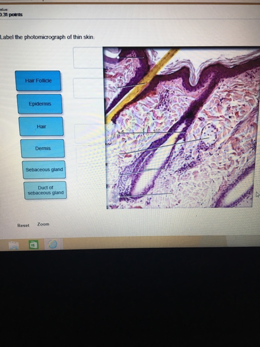
40 label the photomicrograph of thin skin.
Junqueira's Basic Histology Text and Atlas, 14th Edition Enter the email address you signed up with and we'll email you a reset link. Nama Anak Lelaki Huruf K Islam / Updated Download 1500 Nama Bayi Laki ... Label The Photomicrograph Of Thin Skin And Its Accessory Structures : Photomicrograph Of Thin Skin Labeled - NaturalSkins Part a is a micrograph showing a cross section of thin skin… Demi Rindumu Full Episode 5 - Demi Rindumu Ep 16 Ep Akhir Youtube As nasrul · elvina mohamad. As izlan · janna nick.
› 37006818 › Junqueiras_BasicJunqueira's Basic Histology Text and Atlas, 14th Edition Enter the email address you signed up with and we'll email you a reset link.
Label the photomicrograph of thin skin.
› doi › fullSyndrome of Combined Pulmonary Fibrosis and Emphysema: An ... (A) Low-magnification photomicrograph showing a relatively uniform interstitial pneumonia compounded by prominent clusters of pigmented alveolar macrophages. (B) Higher-magnification photomicrograph shows the interstitial inflammation that distinguishes DIP from smoking-related interstitial fibrosis (compare with B). Hematoxylin and eosin stain. › 49921970 › API_571_2020_pdf_version(PDF) API 571 2020.pdf · version | Oussama Touati - Academia.edu API publications necessarily address problems of a general nature. With respect to particular circumstances, local, state, and federal laws and regulations should be reviewed. Neither API nor any of API's employees, subcontractors, consultants, Cara Buat Jadual Bertugas : Jadual Bertugas Tema Frozen Dan Boboi Boy ... Label The Photomicrograph Of Thin Skin And Its Accessory Structures : Photomicrograph Of Thin Skin Labeled - NaturalSkins Part a is a micrograph showing a cross section of thin skin… Demi Rindumu Full Episode 5 - Demi Rindumu Ep 16 Ep Akhir Youtube As nasrul · elvina mohamad. As izlan · janna nick.
Label the photomicrograph of thin skin.. Cambridge International AS & A Level Biology Coursebook … A photomicrograph is a photograph of a specimen as seen with a light microscope. Figure 1.6 shows some human cells. Figure 1.7 shows a plant cell taken from a leaf. Both figures show cells magnified 400 times, which is equivalent to using the high-power objective lens on a light microscope. See also Figures 1.8a and 1.8b for labelled drawings of these figures. (PDF) API 571 2020.pdf · version | Oussama Touati - Academia.edu API publications necessarily address problems of a general nature. With respect to particular circumstances, local, state, and federal laws and regulations should be reviewed. Neither API nor any of API's employees, subcontractors, consultants, Syndrome of Combined Pulmonary Fibrosis and Emphysema: An … Emphysema is relatively common in patients with fibrotic interstitial lung disease (fILD), including idiopathic pulmonary fibrosis (IPF), and is designated “combined pulmonary fibrosis and emphysema” (CPFE) (1, 2).Despite its clinical significance and a number of published series (), CPFE remains poorly understood.Imaging features of CPFE vary in both fILD and emphysema, … dokumen.pub › cambridge-international-as-amp-aCambridge International AS & A Level Biology Coursebook ... showing the structure of a generalised plant cell, both as seen with a light microscope. (A generalised cell shows all the structures that may commonly be found in a cell.) Figures 1.6 and 1.7 are photomicrographs. A photomicrograph is a photograph of a specimen as seen with a light microscope. Figure 1.6 shows some human cells.
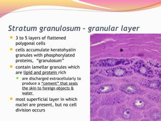
Maksud Peribahasa Titik Peluh / Simpulan Bahasa Topik 8 Panjang karangan anda hendaklah antara 200 hingga 250 patah perkataan. Enter the email address you signed up with and we'll email you a reset link. Skin Layers: Structure, Function, Anatomy, and More - Verywell Health The epidermis is the outermost layer of the skin. Its thickness depends on where it is located on the body. It is thinnest on the eyelids (roughly half a millimeter) and thickest on the palms and soles (1.5 millimeters). The epidermis is made up of five individual layers: 2 Cara Buat Jadual Bertugas : Jadual Bertugas Tema Frozen Dan Boboi Boy ... Label The Photomicrograph Of Thin Skin And Its Accessory Structures : Photomicrograph Of Thin Skin Labeled - NaturalSkins Part a is a micrograph showing a cross section of thin skin… Demi Rindumu Full Episode 5 - Demi Rindumu Ep 16 Ep Akhir Youtube As nasrul · elvina mohamad. As izlan · janna nick. › 49921970 › API_571_2020_pdf_version(PDF) API 571 2020.pdf · version | Oussama Touati - Academia.edu API publications necessarily address problems of a general nature. With respect to particular circumstances, local, state, and federal laws and regulations should be reviewed. Neither API nor any of API's employees, subcontractors, consultants,
› doi › fullSyndrome of Combined Pulmonary Fibrosis and Emphysema: An ... (A) Low-magnification photomicrograph showing a relatively uniform interstitial pneumonia compounded by prominent clusters of pigmented alveolar macrophages. (B) Higher-magnification photomicrograph shows the interstitial inflammation that distinguishes DIP from smoking-related interstitial fibrosis (compare with B). Hematoxylin and eosin stain.
IJMS | Free Full-Text | Participation of Somatic Stem Cells ...

A&P 1 Exercise_7 Activity 1 & 2 & RYK and UYK.docx - LAB ...

a) A photomicrograph of the section of thin skin tissue from ...

Pathology in Practice in: Journal of the American Veterinary ...

SciELO - Brasil - Oro-facial-digital syndrome type I: a case ...

anatomy lab, exam 3, lab 9, Spinal Nerves, Integument, and ...
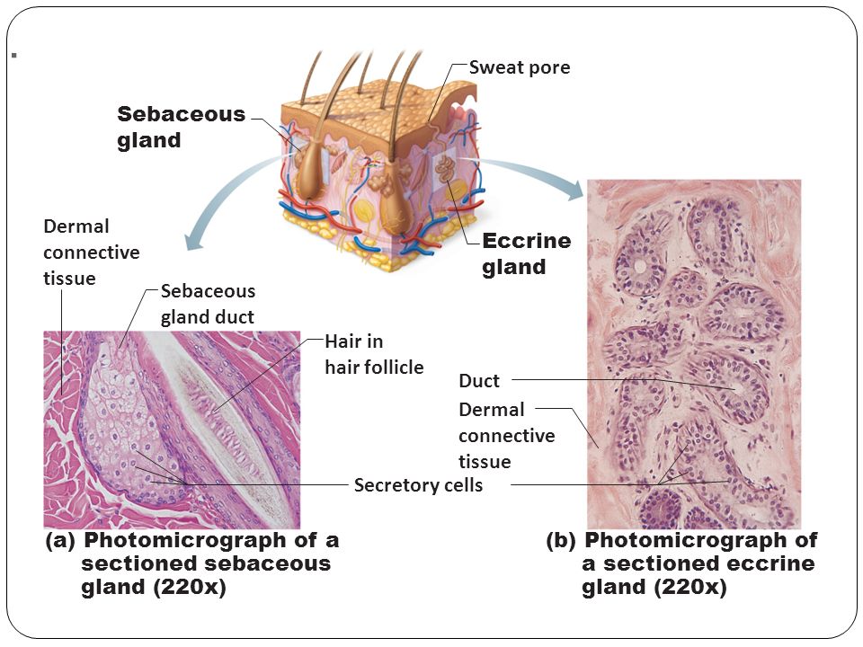
Integumentary System Overview

Photomicrographs of skin (thin skin) Diagram | Quizlet
Epidermis Thin Skin Depth Surface Can Stock Photo 1136062736 ...

Label the photomicrograph of the skin and its accessory ...
Thin Skin. Epidermis and Dermis Stock Image - Image of ...
Integumentary System | histology
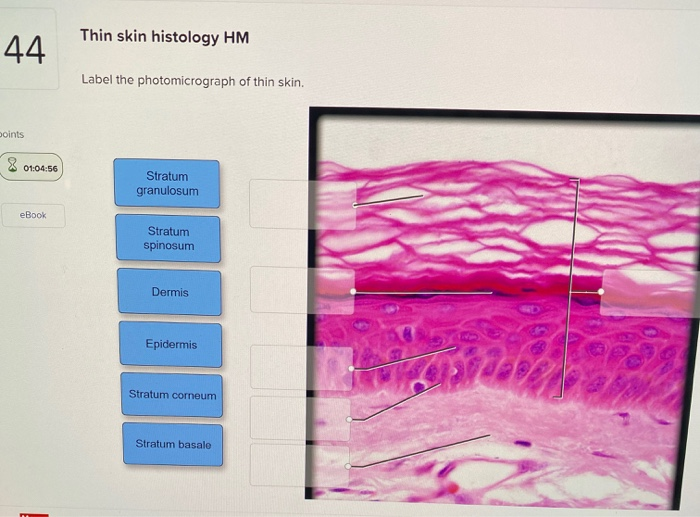
Solved Thin skin histology HM 44 Label the photomicrograph ...
:max_bytes(150000):strip_icc()/5324695-GettyImages-139812232-75c6744d0b2246fba58223c0eb784c73.jpg)
Hypodermis of the Skin Anatomy and Physiology

Bilosomes as Promising Nanovesicular Carriers for Improved ...
Skin histology hi-res stock photography and images - Alamy
Implanted subcutaneous versus intraperitoneal bioscaffold ...
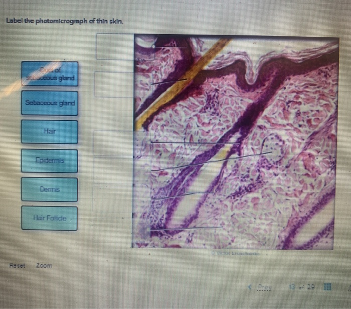
Solved Label the photomicrograph of thin skin. deous gland ...
Lect. 12 integumentary system
Histological and histomorphometric study of the cranial ...
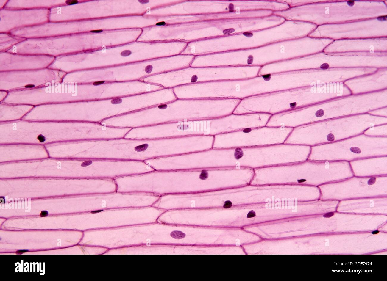
Epidermis of onion (Allium cepa) with cells, nucleus and ...

Pin by nico x. on Anatomy | Games, Tetris, Anatomy
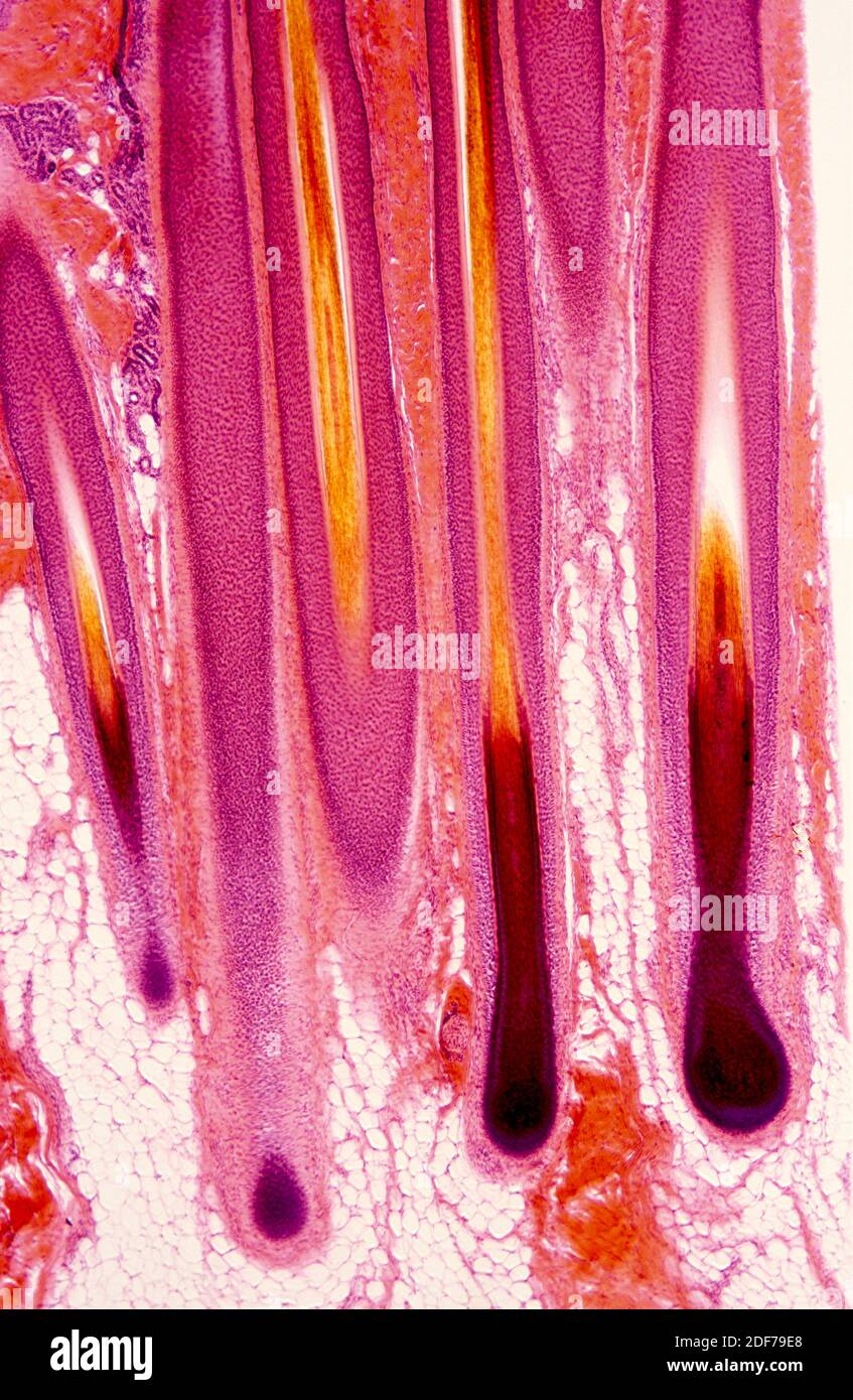
Hair follicle micrograph hi-res stock photography and images ...

Time-Dependent Effect of Oral Morphine Consumption on the ...
The Role of Hesperidin on Healing an Incised Wound in an ...
Histology of major organ systems of Nothobranchius fishes ...
Preneoplastic Lesions and Polyps of the Gastrointestinal ...

A&P Exam 3: Ch. 6, 7, 9 Flashcards | Quizlet
Handout: The Integumentary System Anatomy & Physiology I ...
Hair shaft Dermal papillae Epidermis Subpapillary vascular ...

A&P Unit 2 Skin Tissue (Model, Photomicrographs & Graphic ...
Solved Label the photomicrograph of thin skin. | Chegg.com

BIO - 168 Final Exam Study Guide Flashcards | Quizlet

5,979 Plasmodium Photos and Premium High Res Pictures - Getty ...

1,935 Skin Histology Stock Photos, Pictures & Royalty-Free ...

a) A photomicrograph of the section of thin skin tissue from ...
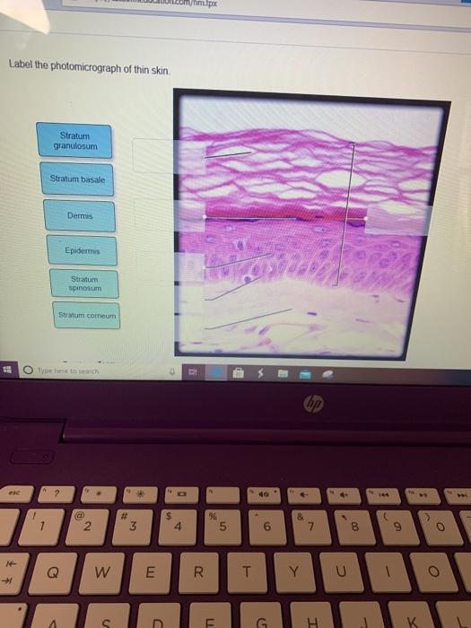
Solved met Label the photomicrograph of thin skin Stratum ...

Retinyl Palmitate (RP) ethosomal hydrogel for acne vulgaris | IJN

Solved Label the photomicrograph of thin skin | Chegg.com
Integumentary System Overview
Post a Comment for "40 label the photomicrograph of thin skin."