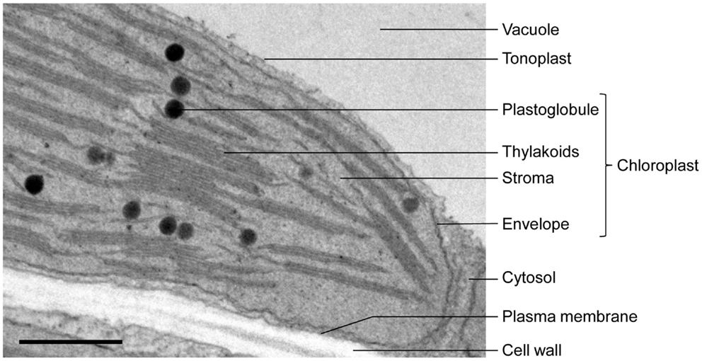
38 transmission electron microscope image of chloroplast labeled
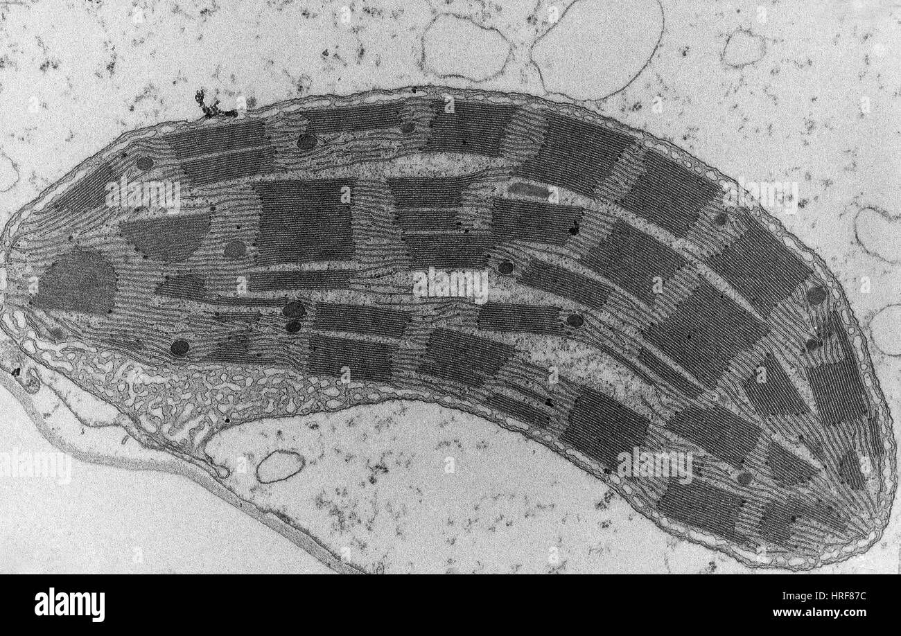
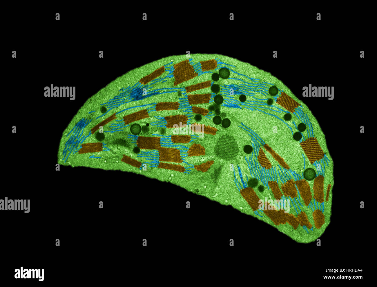
Electron Microscopes Explained: From Physics to Images An electron microscope is a highly advanced microscope that, depending on the type of electron microscope, blasts electrons through a specimen, excites electrons that make up the specimen, or maps the tunneling of electrons through a specimen and reconstructs the feedback from these methods to form an image. The ability of these microscopes to help us visualize specimens that are smaller than ... Chloroplast Micrograph Stock Photos and Images - Alamy Transmission electron micrograph of a chloroplast ID: CXWTYG (RF) Tomato Chloroplast ID: HRF83G (RM) Leaf epidermis, light micrograph ID: 2GAFY49 (RF) Micrasterias truncata.Chlorophyta.Algae.Seaweed.Optic micrsocopy. ID: MTEHMP (RF) Chloroplast TEM ID: HRF5RN (RM) Spirogyra algae, light micrograph ID: 2EA0TJN (RF)
Chloroplast Photos and Premium High Res Pictures - Getty Images chloroplast micrograph chloroplast structure 4,396 Chloroplast Premium High Res Photos Browse 4,396 chloroplast stock photos and images available, or search for chloroplast micrograph or chloroplast structure to find more great stock photos and pictures. NEXT

Transmission electron microscope image of chloroplast labeled
microbenotes.com › under-the-microscopeAmazing 27 Things Under The Microscope With Diagrams A scanning transmission electron microscope (STEM) is often used to observe crystals or compounds that reveal the atoms present inside the compounds with some electrons being used to identify atoms of a particular element through the microscope. The structure of an atom is visible with these microscopes. Electron Microscope- Definition, Principle, Types, Uses, Labeled Diagram There are two types of electron microscopes, with different operating styles: 1. Transmission Electron Microscope (TEM) The transmission electron microscope is used to view thin specimens through which electrons can pass generating a projection image. The TEM is analogous in many ways to the conventional (compound) light microscope. What are the labels of the transmission electronic microscope image of ... Transfer RNA (tRNA) precursors undergo endoribonucleolytic processing of their 5' and 3' ends. 5' cleavage of the precursor transcript is performed by ribonuclease P (RNase P). While in most organisms RNase P is a ribonucleoprotein that harbors a catalytically active RNA component, human mitochondria and the chloroplasts (plastids) and mitochondria
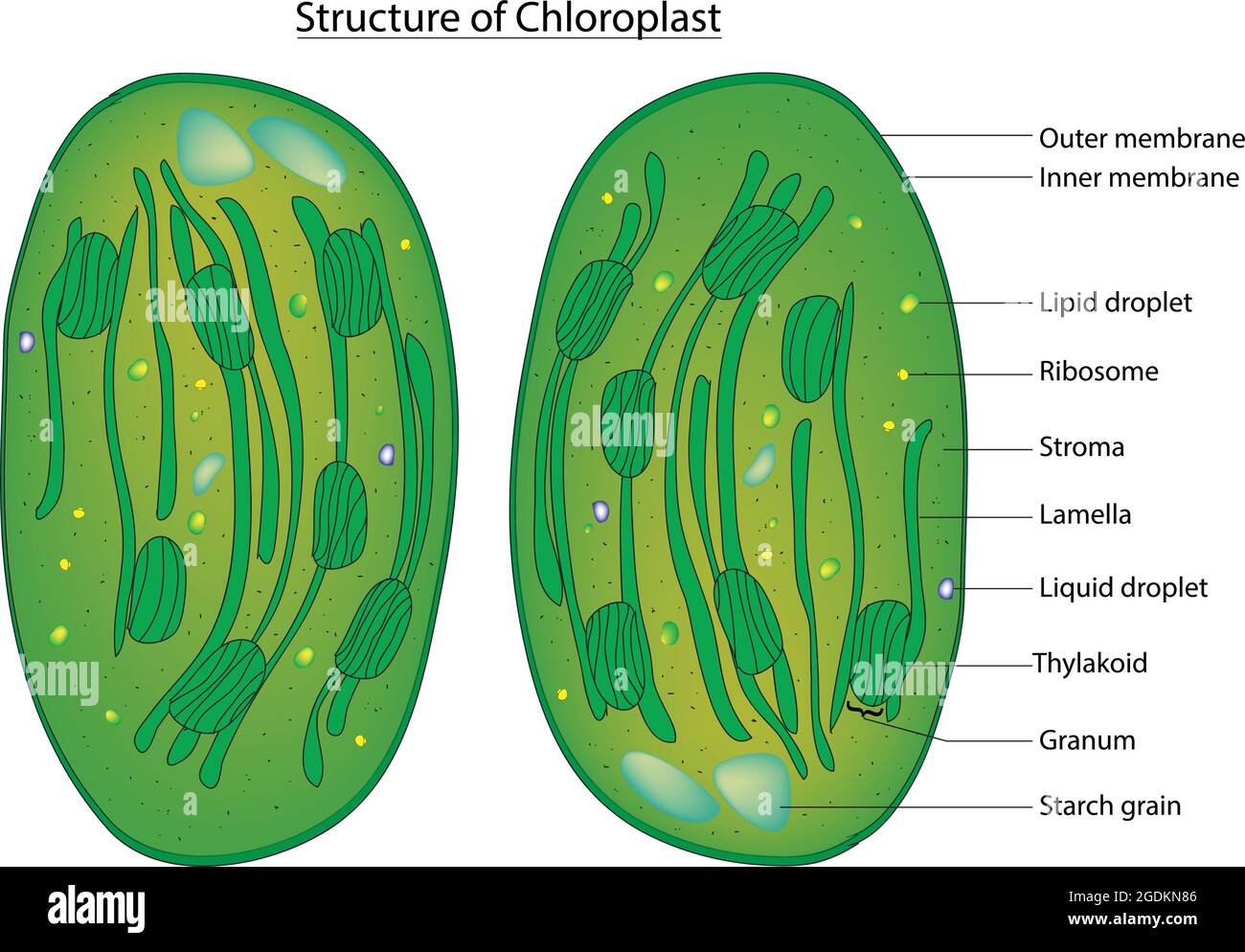
Transmission electron microscope image of chloroplast labeled. transmission electron microscope | instrument | Britannica transmission electron microscope (TEM), type of electron microscope that has three essential systems: (1) an electron gun, which produces the electron beam, and the condenser system, which focuses the beam onto the object, (2) the image-producing system, consisting of the objective lens, movable specimen stage, and intermediate and projector len... Leaf chloroplast - ru Chloroplasts are abundant in the sponge parenchyma of leaves. With field-emission scanning electron microscopy (FESEM) and transmission electron microscopy (TEM) very clear details of the structure of chroroplasts can be observed. FESEM views of sponge parenchyma with chloroplasts Chloroplast High Resolution Stock Photography and Images - Alamy Huge collection, amazing choice, 100+ million high quality, affordable RF and RM images. No need to register, buy now! Lightboxes ; 0 Cart; Account; ... Transmission electron microscope image of a thin section cut from ... Detailed diagram of chloroplast, chloroplast structure, labeled diagram anatomy of the chloroplast with thylakoids and ... Transmission Electron Microscope (With Diagram) - Biology Discussion Finally, the electrons are focused by an electromagnetic projector lens (instead of an ocular lens as in a light microscope) on a screen or photographic plate. The final image in a TEM is known as transmission electron micrograph. The salts of some heavy metals, e.g., lead; osmium, tungsten and uranium are often used for staining.
Chloroplast - Wikipedia A chloroplast / ˈ k l ɔːr ə ˌ p l æ s t,-p l ɑː s t / is a type of membrane-bound organelle known as a plastid that conducts photosynthesis mostly in plant and algal cells.The photosynthetic pigment chlorophyll captures the energy from sunlight, converts it, and stores it in the energy-storage molecules ATP and NADPH while freeing oxygen from water in the cells. The ATP and NADPH is ... Label This Transmission Electron Micrograph : TEM of chloroplast from ... Subset of labeled images and transfer labels to the entire image corpus. Molecular labeling for correlative microscopy: Transmission electron microscopy (tem) is a microscopy technique in which a beam of electrons is transmitted through a specimen to form an image. Label the transmission electron micrograph of the nucleus. Semi-automatic organelle detection on transmission electron microscopic ... (A) TEM image used for detection of etioplasts (inset). Image resolution: 2000 × 2000 pixels. (B) Results of semi-automatic detection. Green rectangles show 'true' etioplasts, which were confirmed by visual inspection; red boxed regions show semi-automatically detected windows. › articles › s41467/022/31751-0Ciliary transition zone proteins coordinate ciliary protein ... Jul 09, 2022 · The transition zone (TZ) of the cilium/flagellum serves as a diffusion barrier that controls the entry/exit of ciliary proteins. Mutations of the TZ proteins disrupt barrier function and lead to ...
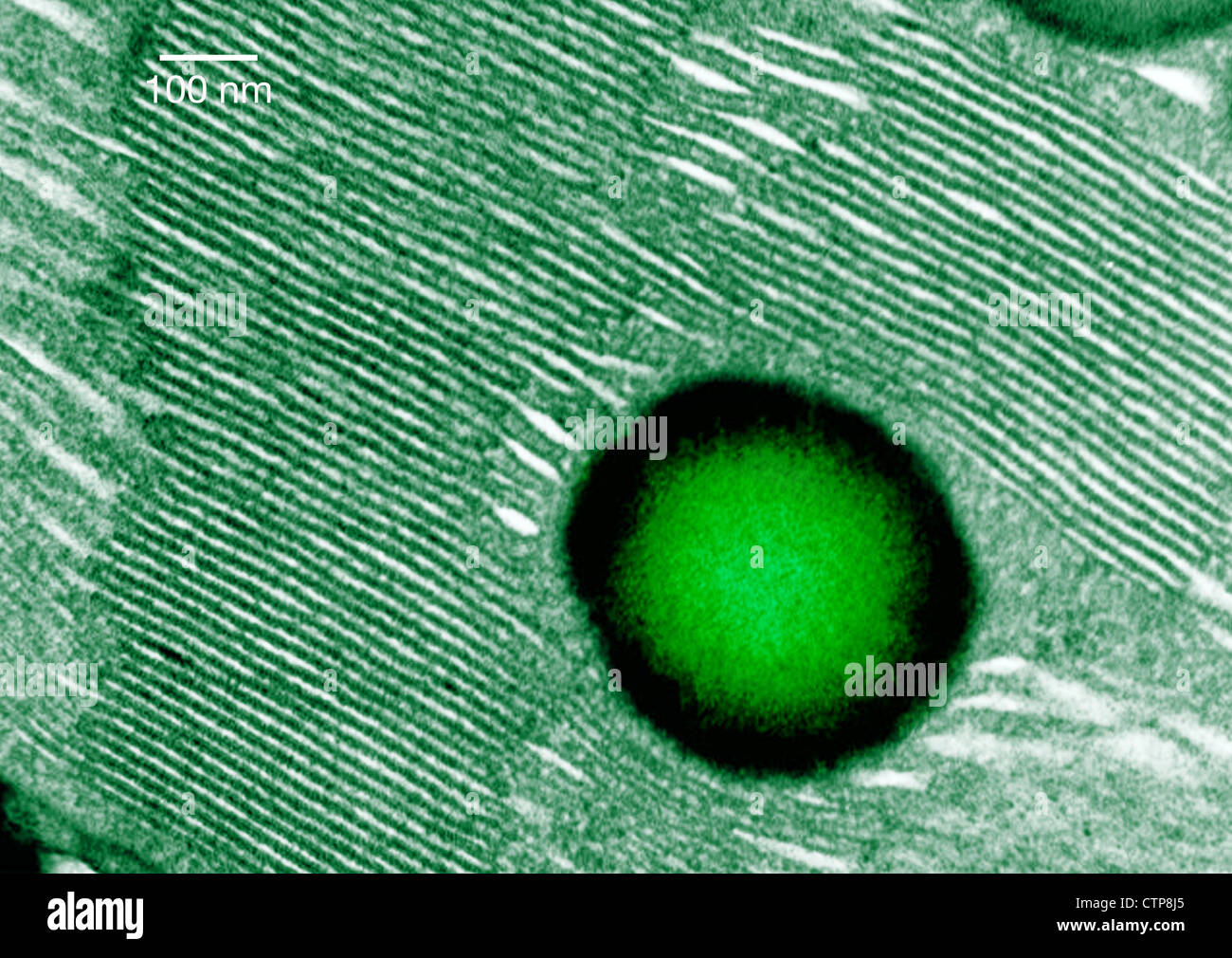
› articles › s41467/022/32109-2Phosphatidylinositol-4-phosphate controls autophagosome ... Jul 28, 2022 · Transmission electron microscopy observations were carried out on a FEI TECNAI Spirit 120 kV electron microscope equipped with an Eagle 4Kx4K CCD camera. Correlation was based on natural landmarks ... Chloroplasts | Photoreceptor Apparata | Algae - Biocyclopedia Transmission electron microscopy image of a chloroplast at higher magnification showing the thylakoid membrane and the eyespot globules (b). (Bar: 1 µm.) FIGURE 2.79 Transmission electron microscopy image of Nannochloropsis sp. in transverse section, showing the chloroplast (a) (Bar: 0.50 mm); chloroplast at higher magnification (b) (Bar: 0.10 ... Transmission Electron Microscope - Quizlet 71 terms. Belinda_Woodard2. Portable and easy to use, Transmission Electron Microscope study sets help you review the information and examples you need to succeed, in the time you have available. Use your time efficiently and maximize your retention of key facts and definitions with study sets created by other students studying Transmission ... Semi-automatic organelle detection on transmission electron ... - Nature Semi-automatic detection of chloroplasts in a TEM image of Arabidopsis thaliana embryo. (A) TEM image used to detect chloroplasts (inset). Image resolution: 2000 × 2000 pixels.

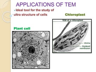
Topic 1.2 Ultrastructure Of Cells - Lessons - Blendspace
Animal Cell Electron Microscope Labelled - Q14 Draw a large diagram of ... A huge resource of stem projects and activities where elementary students apply their knowledge of the major differences between plant and animal scanning electron microscope. Source: images.fineartamerica.com. Using an electron microscope, the electrons can be used to form resolved images of cellular structures of about 3 nm in size.
Tem chloroplast hi-res stock photography and images - Alamy
Transmission Electron Microscope (TEM)- Definition, Principle, Images The working principle of the Transmission Electron Microscope (TEM) is similar to the light microscope. The major difference is that light microscopes use light rays to focus and produce an image while the TEM uses a beam of electrons to focus on the specimen, to produce an image.

DUE TODAY: SYLLABUS/HONOR CODE SIGNED - ppt download
rsscience.com › ribosomesRibosome - protein factory - definition, function, structure ... Ribosomes under an electron microscope. A transmission electron microscope (TEM) is the best tool to observe ribosomes. [In this figure] An electron microscope image of the rough endoplasmic reticulum. The dark spots are ribosomes.
Electron microscope ppt
The Transmission Electron Microscope | CCBER - UC Santa Barbara Transmission electron microscopes (TEM) are microscopes that use a particle beam of electrons to visualize specimens and generate a highly-magnified image. TEMs can magnify objects up to 2 million times. In order to get a better idea of just how small that is, think of how small a cell is.
Thylakoids hi-res stock photography and images - Alamy
pubs.acs.org › toc › ancac3ACS Nano | Vol 16, No 8 Magnetoresistance is an effective probe to detect the coupling between the spin configuration (represented by the gyroscopes) and the charge transport (represented by different colors) and reflect the interaction between different spins of a layered magnetic semiconductor. View the article.

Electron micrograph of isolated chloroplasts with the major ...
Three‐dimensional ultrastructure of chloroplast pockets formed under ... Traced and three-dimensional images of a chloroplast with an open type pocket. (a) Part of the traced serial transmission electron microscopy images of a chloroplast with an open type pocket. The number in each photograph indicates the order of serial sections. Green and purple indicate a chloroplast and lipid body, respectively.

The ANGULATA7 gene encodes a DnaJ‐like zinc finger‐domain ...
Plastids Microscope - frontiers trehalose lipid biosurfactant reduces ... Plastids Microscope - frontiers trehalose lipid biosurfactant reduces adhesion of microbial, sieve tube structure, chloroplast under microscope labeled micropedia, chloroplast sem stock image c001 2430 science photo library, arabidopsis plants in the glass tube, Home Plastids Microscope Plastids Microscope Andrea Saturday, August 20, 2022

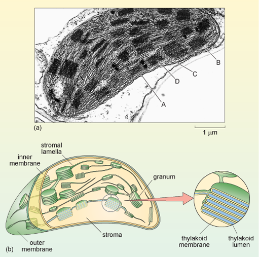
1 Chloroplast. (a) Electron micrograph of a chloroplast in a ...
Chloroplasts - Definition, Structure, Function and Microscopy Microscopy. To view chloroplasts under the microscope, students can use toluidine blue stain to prepare a wet mount. This simply involves the following simple steps: Place a plant sample onto drop of water on a clean glass slide; Using a dropper, add a drop of the stain (toluidine blue) on the sample and allow to stand for about a minute
A tour of the cell: View as single page
Assignment 6, page 2 - North Carolina State University Study this transmission electron micrograph of a spinach leaf cell, locate a chloroplast and capture the image for labeling. The micrograph is displayed as if using a "virtual electron microscope", so you will need to magnify the image and move to a region that contains the clearest view of chloroplast internal structures.
Chloroplast - Wikipedia
› books › NBK26880Looking at the Structure of Cells in the Microscope ... Images of Surfaces Can Be Obtained by Scanning Electron Microscopy. A scanning electron microscope (SEM) directly produces an image of the three-dimensional structure of the surface of a specimen. The SEM is usually a smaller, simpler, and cheaper device than a transmission electron microscope.
Micrograph chloroplast hi-res stock photography and images ...
Transmission electron microscope images of chloroplasts in WT and asl2 ... Download scientific diagram | Transmission electron microscope images of chloroplasts in WT and asl2 mutant. A: Wild-type cell. B: asl2 mutant cell. C: Intact chloroplast in the wild-type cell. D ...

Thin section electron micrograph of a young tobacco ...
Assignment 6, page 1 View this transmission electron micrograph of a plant cell, locate a chloroplast and capture the image for labeling. The micrograph is displayed as if using a "virtual electron microscope", so you will need to magnify the image and move to a region that contains the clearest view of chloroplast internal structures.
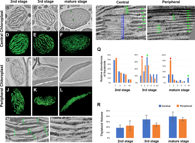
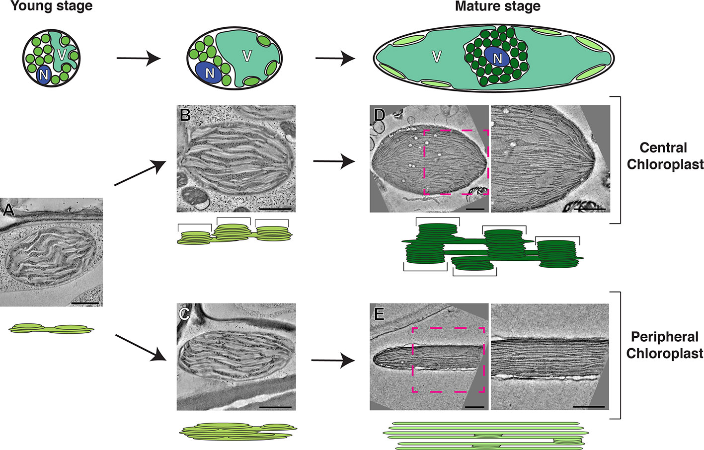
Electron Tomography Analysis of Thylakoid Assembly and ...
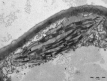
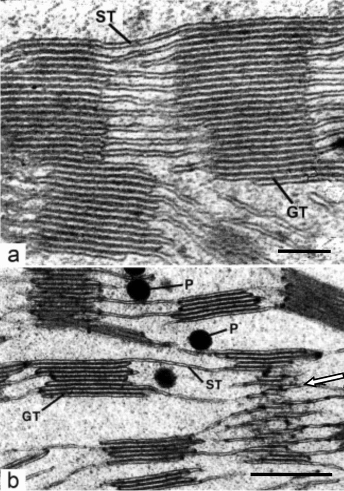
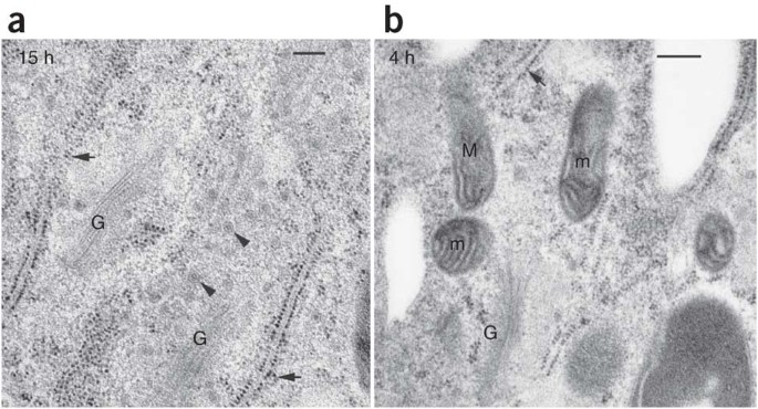
Scanning electron microscopy of chloroplast ultrastructure Transmission electron micrograph of an Aucuba japonica chloroplast showing the cell wall (CW), chloroplast envelope (arrows), grana (G) and plastoglobuli (P). x 15,960 Fig. 3. Scanning electron micrograph of a fractured chloroplast showing incomplete extraction of the stroma after cytoplasmic maceration for 48 hr. x 11,400. Fig. 4.
A tour of the cell: View as single page
Transmission electron microscopic images of chloroplasts and ... Transmission electron microscopic images of chloroplasts and mitochondria in 15-day-old leaves from PRORP1 RNAi mutants and wild-type plants. (A, B) Ultrastructure of chloroplasts and mitochondria...

1.2 Skill: Interpretation of electron micrographs - YouTube
Transmission electron microscopy - Wikipedia Transmission electron microscopy ( TEM) is a microscopy technique in which a beam of electrons is transmitted through a specimen to form an image. The specimen is most often an ultrathin section less than 100 nm thick or a suspension on a grid.

TEM of chloroplasts in a leaf cell - Stock Image - B110/0033 ...
› science › articleProtonated carbon nitride elicits microalgae for water ... Aug 15, 2022 · X-ray photoelectron spectroscopy (XPS) was used to fundamentally understand the interfacial interplay between P-C 3 N 4 and algae. According to the deconvolution results of high-resolution N 1s spectra (), the content of NH x groups increased from 15.5% of pristine g-C 3 N 4 to 19.8% of P-C 3 N 4, indicating the occurence of protonation by acid treatment (Zhou et al., 2015; Du et al., 2015).

Assignment 6, page 2
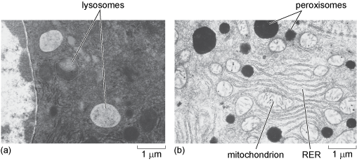
Labeling the Cell Flashcards | Quizlet Label the transmission electron micrograph of the nucleus. membrane bound organelles golgi apparatus, mitochondrion, lysosome, peroxisome, rough endoplasmic reticulum nonmembrane bound organelles ribosomes, centrosome, proteasomes cytoskeleton includes microfilaments, intermediate filaments, microtubules Identify the highlighted structures
30 Label The Transmission Electron Microscope Image Of A ...
What are the labels of the transmission electronic microscope image of ... Transfer RNA (tRNA) precursors undergo endoribonucleolytic processing of their 5' and 3' ends. 5' cleavage of the precursor transcript is performed by ribonuclease P (RNase P). While in most organisms RNase P is a ribonucleoprotein that harbors a catalytically active RNA component, human mitochondria and the chloroplasts (plastids) and mitochondria
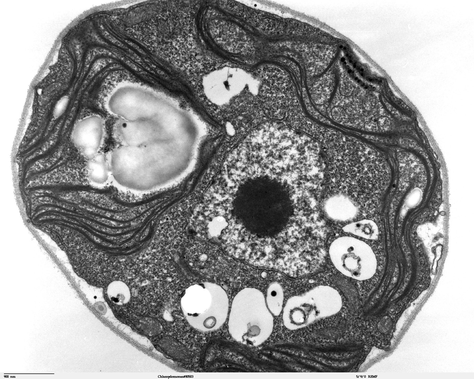
File:Chlamydomonas TEM 07.jpg - Wikimedia Commons
Electron Microscope- Definition, Principle, Types, Uses, Labeled Diagram There are two types of electron microscopes, with different operating styles: 1. Transmission Electron Microscope (TEM) The transmission electron microscope is used to view thin specimens through which electrons can pass generating a projection image. The TEM is analogous in many ways to the conventional (compound) light microscope.
Chloroplast Ultrastructure in Mutant Strains of Chlamydomonas ...
microbenotes.com › under-the-microscopeAmazing 27 Things Under The Microscope With Diagrams A scanning transmission electron microscope (STEM) is often used to observe crystals or compounds that reveal the atoms present inside the compounds with some electrons being used to identify atoms of a particular element through the microscope. The structure of an atom is visible with these microscopes.
A Thylakoid Membrane Protein Functions Synergistically with ...
A brief history of how microscopic studies led to the ...

Cell Micrographs | BioNinja

Cell Micrographs | BioNinja
Nuclear envelope. TEM stock image. Image of micrograph ...
Chloroplast tem hi-res stock photography and images - Alamy
What is a diagram of a plant and animal cell under an ...
Chloroplast tem hi-res stock photography and images - Alamy

Chloroplast - Wikipedia

Thin section electron micrograph of a chloroplast from wild ...

File:Chloroplast in leaf of Anemone sp TEM 30000x.png ...
Frontiers | Electron Microscopy Views of Dimorphic ...
Is chloroplast visible under a light microscope? - Quora
Preparation of plant cells for transmission electron ...
![PDF] The fine structure of chloroplasts and pyrenoids in some ...](https://d3i71xaburhd42.cloudfront.net/6018a6b31f239af4b85d04769542f9e4f73a0e85/10-Figure3-1.png)
PDF] The fine structure of chloroplasts and pyrenoids in some ...
A brief history of how microscopic studies led to the ...

Draw a labelled diagram of chloroplast as seen under an ...
Frontiers | When Proteomics Reveals Unsuspected Roles: The ...
Post a Comment for "38 transmission electron microscope image of chloroplast labeled"