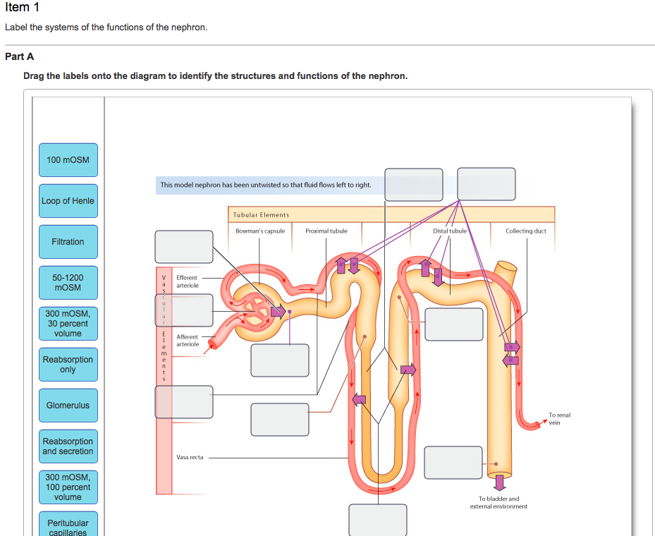
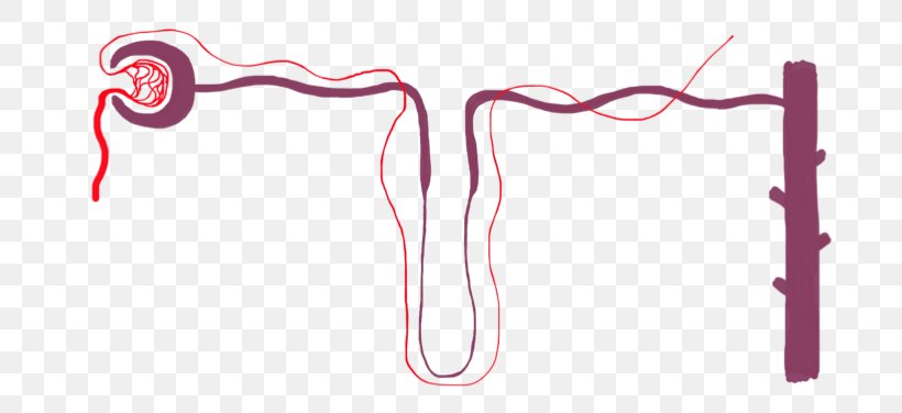
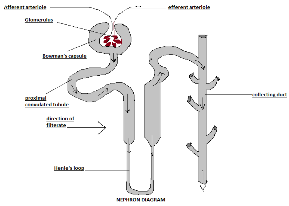
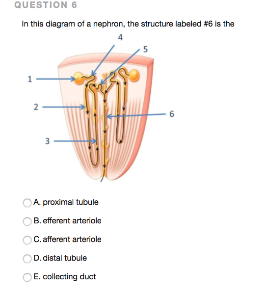
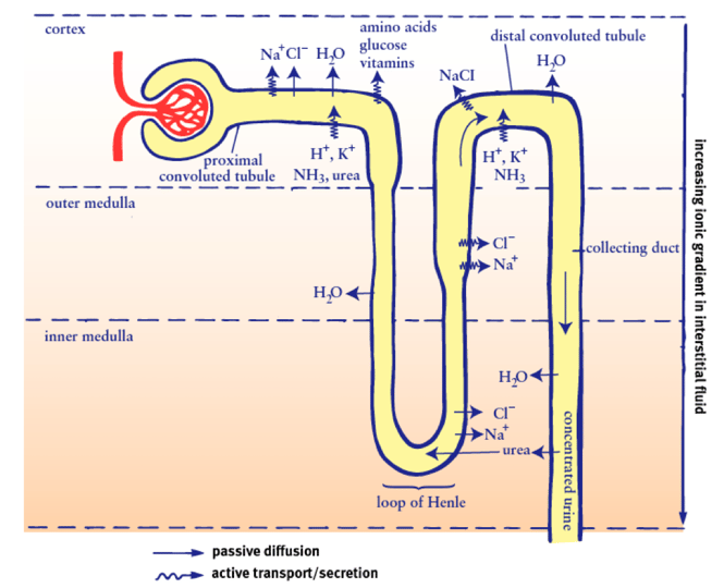
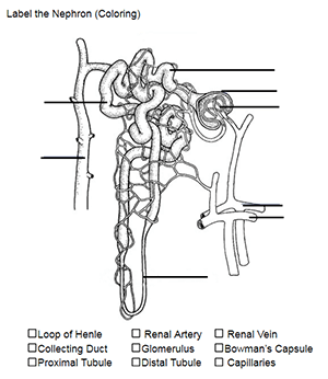
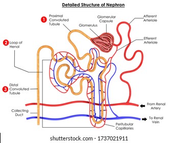
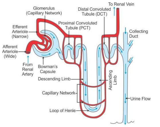
44 labeled diagram of the nephron
Structure And Function Of Body 13th Edition - safss.msu.edu Anatomy and Physiology Structure & Function of the Body Anatomy & ... Nephron Structure and functions Structure And Function Of Body Fun facts. The human body contains nearly 100 trillion cells. There are at least 10 times ... Pituitary Gland: Anatomy, Function, Diagram, Conditions ... Page 28/31. Bookmark File PDF Kidney Structures and Functions Explained (with Picture and Video) The nephron consists of a renal corpuscle, a tubule, and a capillary network that originates from the small cortical arteries. Each renal corpuscle is composed of a glomerulus (a network of capillaries) and a Bowman's capsule (the cup-shaped chamber that surrounds it.
Anatomy And Physiology Archive | August 08, 2022 | Chegg.com Identify the main regions of the kidney Draw a labeled diagram of a nephron and of the blood supply of the nephron Summarize the ultrastructural features of different parts of the nephron. 1 answer 1.Illustrate the mechanism how hypoxia destroys the cell membrane. 2. How does the body repair a bone after the fracture occurs? 3.What would happen ...

Labeled diagram of the nephron
› doi › 10A reference tissue atlas for the human kidney | Science Advances Jun 08, 2022 · Some nephron segments, such as the loop of Henle and the collecting duct, contain multiple cell types with different reabsorption mechanisms (36, 40); hence, we mostly focus on cell type–specific transport mechanisms . We define mRNA levels mapping to transporters involved in blood-to-lumen transport (table S12) as negative to account for the ... Understanding an ECG | ECG Interpretation | Geeky Medics An ECG electrode is a conductive pad that is attached to the skin to record electrical activity. An ECG lead is a graphical representation of the heart's electrical activity which is calculated by analysing data from several ECG electrodes. A 12-lead ECG records 12 leads, producing 12 separate graphs on a piece of ECG paper. art-labeling activity: the structure of the digestive tract A generalized nephron and collecting system. The question here wants us to identify the part of the desk digestive system that includes the following. This consists of 9 worksheets for teaching the digestive system for high school anatomy physiology. Anatomy and Physiology questions and answers. Figure 2331a 2 of 2. Figure 2331a 2 of 2.
Labeled diagram of the nephron. › structures › structureStructure of the Kidney (With Diagram) | Organs | Human ... Nephron: Nephron is the basic unit of kidney. The minute structure of the kidney is composed of a number of nephrons. Each human kidney possesses about 1 -2 millions of nephrons. Each nephron is made up of two main parts: (1) Malpighian Body, (2) Renal tubule. (C) Blood Vessels: The two important blood vessels of the kidney are: (1) Renal Artery Label Kidney Diagram - anatomy physiology and pathophysiology, free ... Label Kidney Diagram. Published by Alice; Sunday, August 14, 2022 Glomerulus: Anatomy, structure and functions. | Kenhub The glomerulus is the main filtering unit of the kidney. It is formed by a network of small blood vessels ( capillaries) enclosed within a sac called the Bowman's capsule. The space inside the capsule that surrounds the glomeruli is known as the Bowman's space. Each glomerulus is located at the beginning of the nephron. art-labeling activity: components and divisions of the pelvis Components And Divisions Of The Pelvis 33 Label The Major Systemic Arteries - Labels For Your Ideas The pelvis is inferior to the abdomen. Posted 24 days ago. Transdtional epithelium of the urinary bladder Part A Drag the labels onto the diagram to identify structures associated with the transitional epithelium of the urinary bladder.
NECO Biology Questions and Answers For 2022/2023 (Theory and ... - Bekeking The questions below are the NECO past questions and answers that will help you in your 2021 NECO Biology Questions. 1. Plants are classified into the following classes except (a)Bryophyte. (b) Coelenterate. (c) pteridophyta. (d) thallophyta. (e) Spermatophyte. 2. The cell wall of plants is rigid due to the presence of? (a) Cambium (b) cellulose Anatomy And Physiology Archive | August 04, 2022 | Chegg.com Anatomy And Physiology Archive: Questions from August 04, 2022. please help answer these Anatomy questions. Select from the options below to fill in the blanks. In the anatomical position, the palmar surface can be observed From the anatomical position, if the hand is supinated, it would mean that the palme. 1 answer. Urinary System Answer Key Use the key letters to identify the diagram of the nephron (and associated renal blood supply) on the left. 1. arcuate artery 2. arcuate vein 3. afferent arteriole 4. collecting duct 5. distal convoluted tubule 6. efferent arteriole 7. glomerular capsule 8. glomerulus 9. interlobar artery 10. interlobar vein 11. Glacier Diagram Labeled - geography extreme landscapes glaciers ... Glacier Diagram Labeled - 16 images - geography space place and pretty wellies glacial movement, landforms and waterways, cold environments subject knowledge pedagogy development, gotbooks,
Glacier Diagram Labeled - geography extreme landscapes july 2012 ... Glacier Diagram Labeled - 14 images - karst landscapes diagram science learning hub, geography extreme landscapes july 2012, geography extreme landscapes glaciers, floodplain landforms geography lessons floodplain, Biology Paper 2 Questions and Answers - Mumias West Pre Mocks 2022 The diagram below illustrates the structure of the kidney nephron. Name the part labeled E. (1 mark) How is the part labeled F adapted to its function? (4 marks) State three physiological mechanisms of controlling the human body temperature during a cold day. (3 marks) Blank Kidney Diagram - label kidney diagram human body anatomy, medical ... Blank Kidney Diagram - 16 images - kidney cross section diagram patient, kidney labelled diagram, kidney diagram, excretory system worksheet wikieducator, What Are the Functions of Nephron? | Med-Health.net The nephron is the basic functional and structural unit found in the kidneys. Its main functions include regulating the concentration of sodium salts and water by filtering the kidney's blood, excreting any excess in the urine and reabsorbing the necessary amounts.
Kidney histology: Nephron, loop of Henle, functions | Kenhub The nephron loop is the U-shaped bend of a nephron which extends through the medulla of the kidney. Histologically, it consists of two parts; thin descending and thin ascending limbs. Both limbs are composed of simple squamous epithelium. The cells have few organelles, little to no microvilli and low secretion abilities.
Nephron Tubules - Human Physiology - 78 Steps Health Trace the course of blood flow through the kidney from the renal artery to the renal vein. 3. Trace the course of tubular fluid from the glomerular capsules to the ureter. 4. Draw a diagram of the tubular component of a nephron. Label the segments and indicate which parts are in the cortex and which are in the medulla. Physiology of the Kidneys
› cell-stem-cell › fulltextA scalable organoid model of human autosomal dominant ... Jul 07, 2022 · We describe a scalable platform to efficiently generate thousands of comparable human kidney organoids. In this, PKD1 and PKD2 mutant organoids displayed robust cystogenesis. We generated a small-molecule screening workflow that identified compounds inhibiting cyst formation and later growth. The platform will facilitate kidney disease modeling and high-throughput drug screens.
en.wikipedia.org › wiki › Proximal_tubuleProximal tubule - Wikipedia Proximal convoluted tubule (pars convolutaThe pars convoluta (Latin "convoluted part") is the initial convoluted portion. [citation needed]In relation to the morphology of the kidney as a whole, the convoluted segments of the proximal tubules are confined entirely to the renal cortex.
Structure Of The Nephron Coloring Worksheet Answers (PDF) - api.it.aie labels to identify structures, reinforcing visual and auditory learning from the text. You can also refer to the text if you are uncertain or need to review an area. Unlabeled line drawings and images from every chapter allow for immediate, thorough review of material - and let you refer to the text's diagrams and Workbook's appendix for answers.
Stages of External Respiration & Gradients - My Paper Support Stages of External Respiration & Gradients Stages of External Respiration & Gradients 1. core concept of flow-down gradients, explain the movement of glucose in and out of the nephron. 2. core concept of homeostasis, explain how the kidneys are involved in controlling fluid osmolarity. 3. Compare the structure and functions of the major regions of the nephron.
› questions-and-answers › describeAnswered: Describe the difference between a… | bartleby Q: The following can be found in the renal cortex except collecting tubules Bowman's capsule nephron. A: The kidneys are bean-shaped excretory organs and it is the main organs of the urinary system as it…
Brigitte Zimmer Human Heart Diagram And Label. 9Th Grade Easy Plant Cell Diagram. Simple Diagram Of A Heart With Labels. Label The Picture Of A Human Digestive System. Labeled Sketch Of A Chloroplast. Internal Female Reproductive System Labelled Diagram. Dna Easy Drawing. Digestive System Diagram Class 10 Easy. Easy Plant Cell And Animal Cell Drawing With Labels.
› en › libraryUrinary system: Organs, anatomy and clinical notes | Kenhub Aug 02, 2022 · The organs of the urinary system are the kidneys, ureters, bladder and urethra. The kidneys perform the filtration functions of the urinary system and create urine, while the remaining organs act as transport tubes or provide temporary urine storage. The anatomy of the urinary system can be seen here in the urinary system diagram.
en.wikipedia.org › wiki › Renal_calyxRenal calyx - Wikipedia The renal calyces are chambers of the kidney through which urine passes. The minor calyces surround the apex of the renal pyramids.Urine formed in the kidney passes through a renal papilla at the apex into the minor calyx; two or three minor calyces converge to form a major calyx, through which urine passes before continuing through the renal pelvis into the ureter
12th Biology Chapter Genetic Basis Of Inheritance ? - careers.mass 12th-biology-chapter-genetic-basis-of-inheritance 3/21 Downloaded from careers.mass.gov on July 31, 2022 by guest increasingly being used for a wide range of non-clinical
Renal Sodium and Water Regulation | Concise Medical Knowledge - Lecturio Renal Sodium and Water Handling. A nephron Nephron The functional units of the kidney, consisting of the glomerulus and the attached tubule. Kidneys: Anatomy is the functional unit of the kidney through which fluid and solutes, including Na +, are filtered, reabsorbed, and secreted.. Glomerulus and proximal tubule Proximal tubule The renal tubule portion that extends from the bowman capsule in ...
Diagram of Human Heart and Blood Circulation in It Four Chambers of the Heart and Blood Circulation. The shape of the human heart is like an upside-down pear, weighing between 7-15 ounces, and is little larger than the size of the fist. It is located between the lungs, in the middle of the chest, behind and slightly to the left of the breast bone. The heart, one of the most significant organs ...
(Solved) - 6. The Diagram Below Of Part Of A Nephron ... - Transtutors The Diagram Below Of Part ...
Prostate: Anatomy, Function, and Treatment - Verywell Health The prostate is an important gland located between the penis and bladder. It sits just to the front of the rectum. The urethra, which carries urine from the bladder out of the body, runs through the center of this walnut-sized organ. The gland's primary function is to secrete fluid that nourishes sperm and keeps it safe. 1.
Epithelial Characteristics Of The Nephron - Medical Physiology All nephron segments, from the glomerulus to the ducts of Bellini, consist of epithelial cells that are joined in a continuous layer by specialized structures called junctional complexes, as illustrated in Fig. 1. Although some epithelia consist of multiple cell layers, a single cell layer forms the epithelium of the nephron.
The Urinary System: Nephron & Urine Formation - Owlcation Structure of the Nephron There are more than a million nephrons packed in the renal cortex of the kidney. The nephron is made up of the glomerulus and a system of tubes. The glomerulus is a network of intertwined capillaries mass. It is enclosed in a cup-shaped structure called the bowman's capsule.
BIOL 203 - Anatomy & Physiology II - Acalog ACMS™ - Montcalm Identify the parts of the urinary system on cadaver, model or diagram. Describe the function of the urinary system organs. Describe the structure of a nephron. Describe the function, control and regulation of the pathway of blood/urine through the urinary system. Describe selected urinary disorders, diseases and their causes.
art-labeling activity: the structure of the digestive tract A generalized nephron and collecting system. The question here wants us to identify the part of the desk digestive system that includes the following. This consists of 9 worksheets for teaching the digestive system for high school anatomy physiology. Anatomy and Physiology questions and answers. Figure 2331a 2 of 2. Figure 2331a 2 of 2.
Understanding an ECG | ECG Interpretation | Geeky Medics An ECG electrode is a conductive pad that is attached to the skin to record electrical activity. An ECG lead is a graphical representation of the heart's electrical activity which is calculated by analysing data from several ECG electrodes. A 12-lead ECG records 12 leads, producing 12 separate graphs on a piece of ECG paper.
› doi › 10A reference tissue atlas for the human kidney | Science Advances Jun 08, 2022 · Some nephron segments, such as the loop of Henle and the collecting duct, contain multiple cell types with different reabsorption mechanisms (36, 40); hence, we mostly focus on cell type–specific transport mechanisms . We define mRNA levels mapping to transporters involved in blood-to-lumen transport (table S12) as negative to account for the ...













Post a Comment for "44 labeled diagram of the nephron"