44 art-labeling activity: structure of muscle tissues
PDF Marieb HA8 chapter 4 - Pearson The word tissue derives from the Old French word meaning "to weave," reflecting the fact that the different tissues are woven together to form the "fabric" of the human body. The four basic types of tissue are epithelial tissue, connective tissue, muscle tissue, and nervous tissue. If a single, broad functional term were assigned to ... Answer correct art based question chapter 4 question - Course Hero ANSWER: Correct Intercalated disks are found between cardiac muscle cells and allow them to communicate and contract in unison. Art-Labeling Activity: Structure of muscle tissues Part A nose external ear intervertebral discs fetal skeleton True False intercalated disks dendrites nuclei striations
Solved Secure https:/ C Lab: Histology Art-labeling | Chegg.com Secure https:/ C Lab: Histology Art-labeling Activity: Structure of muscle tissues 102091378 Part A Drag the appropriate labels to their respective targets tissue Smooth muSECM) (cardiac tissue (ECM) (skeletal Nucleus issue Type here to search.
Art-labeling activity: structure of muscle tissues
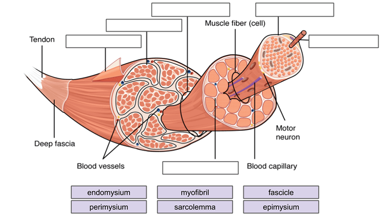
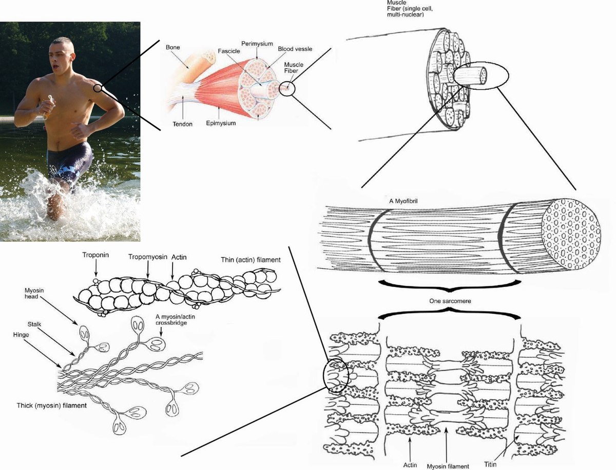
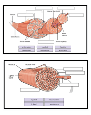
art-labeling activity: the cell and its organelles - Blogger Smooth muscle cells are also relatively large but are long and spindle shaped. Take a toothpick and gently scrape some cells on the inside of the cheek. State a function of each organelle. Organelles are the functional parts of cellsthey are inside the cells in the cytoplasm. Sketch a model of a cell and label its parts. Answered: Art-labeling Activity: Structural… | bartleby Art-labeling Activity: Structural organization of skeletal muscle Reset Epimysium Muscle fascicle Endomysium Perimysium Nerve Muscle fibers Blood vessels Tendon Muscle fiber (cell) PDF In this chapter, you will learn that - Pearson Electron micrograph of a bundle of skeletal muscle fibers wrapped in connective tissue. < 278 Muscles and 9 Muscle Tissue Muscles use actin and myosin molecules to convert the energy of ATP into force In this chapter, you will learn that and finally, exploring Developmental Aspects of Muscles and and and and and 9.2 Gross and microscopic anatomy
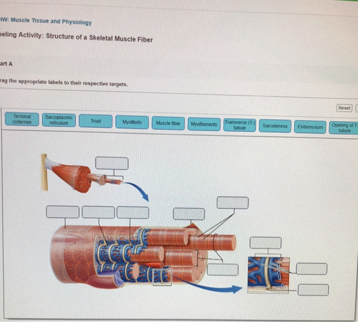
Art-labeling activity: structure of muscle tissues. [Solved] Art-Labeling Activity: | Course Hero The image attached below is a sketch of the layers of the skin. There are 5 layers of the epidermis, which are categorized as follows (from superficial to deep): Stratum corneum: outer layer of the epidermis. Consists of dead (anuclear), keratin-filled cells. This layer is constantly being sloughed off. Stratum lucidum: thin, translucent layer. Art-labeling Activity: The Structure of a Skeletal Muscle Fiber Start studying Art-labeling Activity: The Structure of a Skeletal Muscle Fiber. Learn vocabulary, terms, and more with flashcards, games, and other study tools. ... Write. Test. PLAY. Match. Created by. BabeRuthless0504. Terms in this set (2) Art-labeling Activity: The Structure of a Skeletal Muscle Fiber... Art-labeling Activity: The Structure ... Art Labeling Activity: Layers Of The Uterine Wall : Solved Reproductive ... You can easily copy the label edit art into the lecture presentations by using either the powerpoint. The uterine wall is made up of three layers of muscle tissue. Surrounded by a layer of connective tissue called the tunica albuginea . Practice on pictures with no labels (including no labels for what type of. Answered: Art-labeling Activity: Structural… | bartleby Solution for Art-labeling Activity: Structural classification of joints Suture Symphysis Gomphosis Synostosis or Synovial Syndesmosis ary Synchondrosis n- DO
exercise 19 review sheet art-labeling activity 1 - kamilahlomeli Correct Exercise 6 Review Sheet Art-labeling Activity 1 Identify the tissues and their components. Gross anatomy of the brain and cranial. Relative size of the muscle. Learn vocabulary terms and more with flashcards games and other study tools. Lab manual review sheets have been integrated to follow each exercise. Art-labeling Activity: Types of Connective Tissue Proper Start studying Art-labeling Activity: Types of Connective Tissue Proper. Learn vocabulary, terms, and more with flashcards, games, and other study tools. Stomach histology: Mucosa, glands and layers | Kenhub The innermost layer of the stomach wall is the gastric mucosa.It is formed by a layer of surface epithelium and an underlying lamina propria and muscularis mucosae. The surface epithelium is a simple columnar epithelium.It lines the inside of the stomach as surface mucous cells and forms numerous tiny invaginations, or gastric pits, which appear as millions of holes all throughout the stomach ... 3: Cells and Tissues - Pearson Art-labeling Activities Use the art-labeling activities to quiz yourself on key anatomical structures in this chapter. Figure 3.4: Structure of the generalized cell
art-labeling activity: figure 9.6 - lineartdrawingswallpaperiphone Label the sides of the designs with their lengths. Figure 97 2 of 2 Art-labeling Activity. Use a computer art program to draw two rectangles whose dimensions are. Uvula hyaline cartilage rings vocal folds true vocal cords. Together with the maxillae they comprise the hard palate. In humans it also forms part of the nose and eye socket. pearson anatomy compact bone diagram Long Bone Diagram Pearson / Art-labeling Activities - Diagram Of Of A leonardbone.blogspot.com. pearson labeling ch06. Bone Tissue Anatomy - Google Search | Compact Bone, Anatomy And . bone compact anatomy diagram physiology microscopic label tissue bones drawing structures study. Microscopic Structure Of Compact Bone Quiz Art Labeling Activity the Major Systemic Arteries April 17th 2019 - Art labeling Activities Art labeling Activity Figure 21 5 The Organization of a Capillary Bed Art labeling Activity Figure 21 25 Major Arteries of the Trunk Practice terms with the Crossword Puzzle Step 3 Take the Practice Test Interactive Physiology with Quizzes. Exercise 10 Review Sheet Art-labeling Activity 2 Use the art-labeling activities to quiz yourself on key anatomical structures in this chapter. Shoulder girdle bone that does not articulate with the axial skeleton. Activity 1 Identifying the Bones of the Skull The bones of the skull Figures 91910 pp. Experts are tested by Chegg as specialists in their subject area.
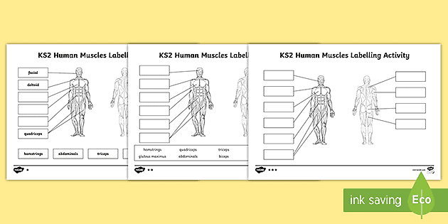
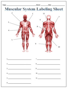
Muscles Labeling - The Biology Corner Part 2 of the slides goes into more detail about how this disease affects the dystrophin in the muscles and how it is inherited as a sex-linked disorder. The activity linked below is a drag and drop activity for students to practice labeling the muscles, there are 6 slides showing images of muscles and fibers and the connective tissue surrounding the fibers (endomysium, perimysium, epimysium).
art-labeling activity: figure 8.3 - gamezombie-danny Review Figure 92 Label Art Activity Figure. Anatomical structure of the shoulder Joint 1 of 2 6 of 37 Part A Drag the appropriate labels to their respective targets Reset Rosat Help Acromion Subscapularis muscle Rotator cuft Tondon of tres minor muscle Tendon or supraspinatus muscle Clavida. 193 Cardiac Cycle. Contrast wavelength and amplitude ...
Solved Art-labeling Activity: Types of Muscle Tissue Reset - Chegg Question: Art-labeling Activity: Types of Muscle Tissue Reset Help Caswelong, ed, and LOCATIONSCombined choti Strations FUNCTION MOVES deti, Smooth Muscle Tissue het Cardac Musce Tissue Cardiac muscle cols and , Skeletal Muscle Tissue LOCATION FUNCTIONS Na intercalated discs Smooth muscle Musdebe LOCATIONS: o Type here to search i. Question.
PDF In this chapter, you will learn that - Pearson Electron micrograph of a bundle of skeletal muscle fibers wrapped in connective tissue. < 278 Muscles and 9 Muscle Tissue Muscles use actin and myosin molecules to convert the energy of ATP into force In this chapter, you will learn that and finally, exploring Developmental Aspects of Muscles and and and and and 9.2 Gross and microscopic anatomy
Answered: Art-labeling Activity: Structural… | bartleby Art-labeling Activity: Structural organization of skeletal muscle Reset Epimysium Muscle fascicle Endomysium Perimysium Nerve Muscle fibers Blood vessels Tendon Muscle fiber (cell)
art-labeling activity: the cell and its organelles - Blogger Smooth muscle cells are also relatively large but are long and spindle shaped. Take a toothpick and gently scrape some cells on the inside of the cheek. State a function of each organelle. Organelles are the functional parts of cellsthey are inside the cells in the cytoplasm. Sketch a model of a cell and label its parts.

:watermark(/images/watermark_only_sm.png,0,0,0):watermark(/images/logo_url_sm.png,-10,-10,0):format(jpeg)/images/anatomy_term/skeletal-muscle/0kdrKFzTosiUmeSgQIFJjQ_Skeletal_muscle_01.png)










Post a Comment for "44 art-labeling activity: structure of muscle tissues"