38 nematocyst diagram
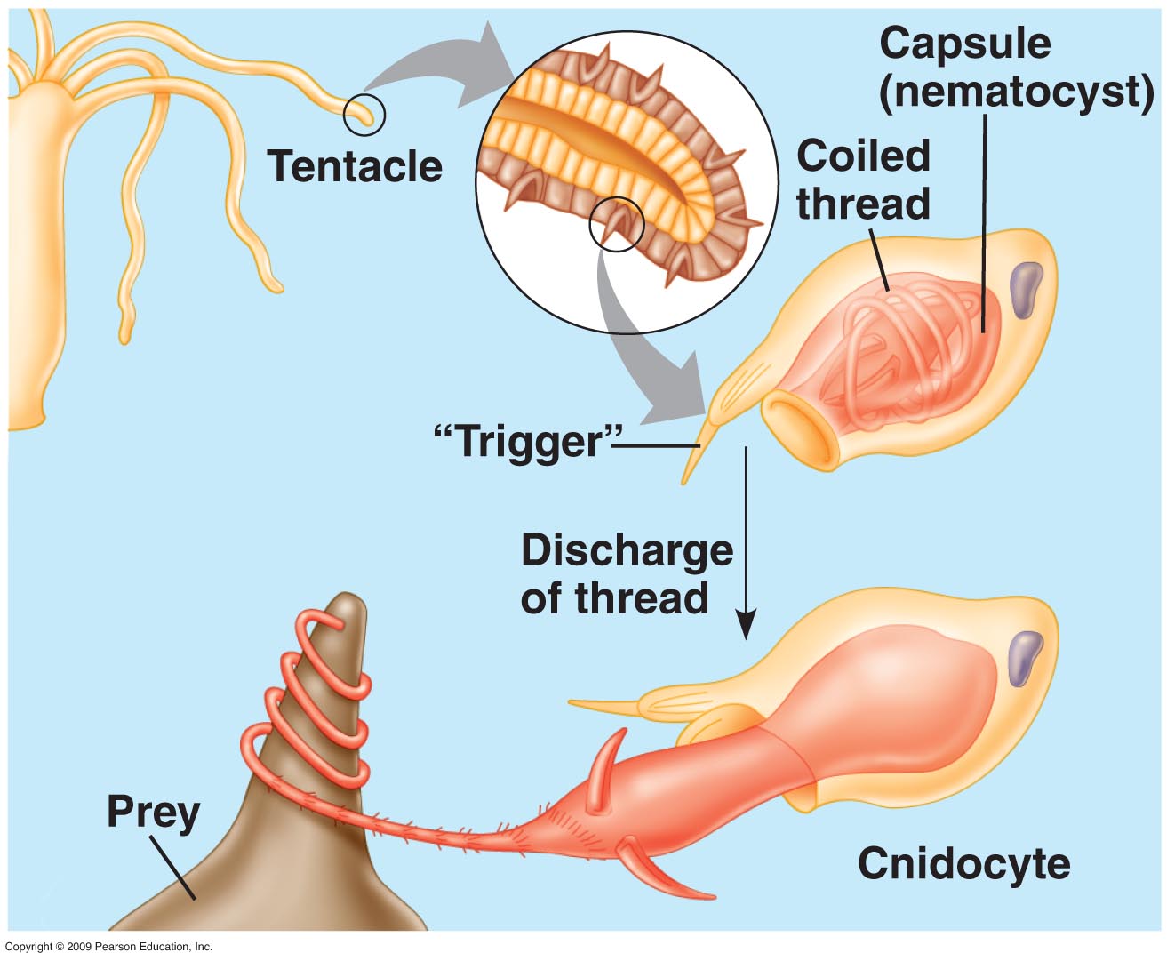
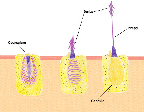
File:Nematocyst discharge.png - Wikimedia Commons Oct 13, 2007 · DescriptionNematocyst discharge.png English: The diagram above shows the anatomy of a nematocystcell and its “firing” sequence, from left to right. On the far left is a nematocyst inside its cellular capsule. The cell’s thread is coiled under pressure and wrapped around a stinging barb. Quantitative Insights into the Contribution of Nematocysts to the ... The nematocyst is a phenotypic novelty in cnidarians, observed in fossils from the Middle Cambrian period [ 1, 8 ]. Each nematocyst consists of a capsule and a coiled tubule through which venom is injected with an ultrafast acceleration of 5 million g [ 9, 10 ].
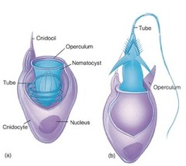
Difference Between Cnidocyte and Nematocyst What is a Nematocyst? It is a subcellular structure or organelle containing extrusive filaments found in a group of unicellular eukaryotes and Cnidarians. It is present inside the cnidocyte and helps it to eject the hair/thread projection as a penetrant, volvent, and glutinant.

Nematocyst diagram
Nematocyst Definition and Examples - Biology Online Dictionary A nematocyst diagram can be seen in Figure 1 below. Figure 1: Diagram of nematocysts in coral. Image Credit: NOAA.gov. In cnidarians, nematocysts are important in the action of locomotion as well as prey capturing and defense mechanisms. These are seen in jellyfish nematocysts and many others. Structure of a nematocyst. cnidocyte. Vector diagram - Shutterstock Structure Nematocyst Cnidocyte Vector Diagram Stock Vector (Royalty Free) 176251196 Download for free See more Popularity score High Usage score High usage Superstar Shutterstock customers love this asset! Item ID: 176251196 Structure of a nematocyst. cnidocyte. Vector diagram Formats EPS 5019 × 3910 pixels • 16.7 × 13 in • DPI 300 • JPG Diagrammatic representations of nematocyst types in Palythoa ... Diagrammatic representations of nematocyst types in Palythoa tuberculosa (the same types are found in Protopalythoa), exploded and unexploded. A,A', Large holotrich (A at one-third scale), scapus....
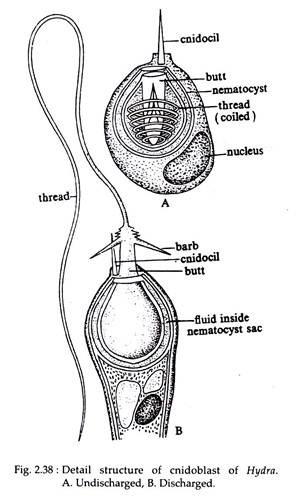
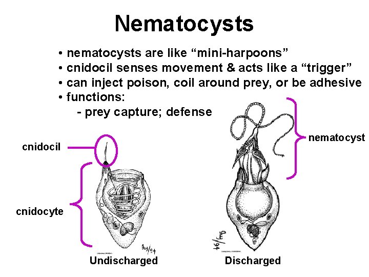
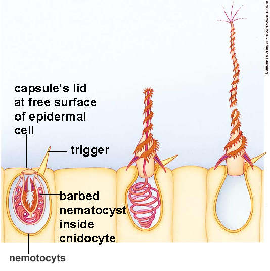
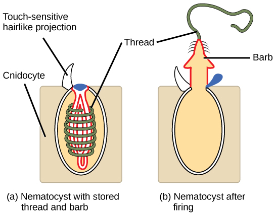
Nematocyst diagram. Nematocysts - The Stinging Cells | Zoology for IAS, IFoS and other ... Nematocysts (Gr. Nema = thread + kystis = bladder) are specialized stinging cells specific to Coelenterates that act as organs of offence and defense. They are also called Cnidae and hence the coelenterates are also called Cnidarians. The cells that produce nematocysts are called nematoblasts. STRUCTURE Nematocysts are cell organelles found in specialized cells called Dynamically Evolving Nematocyst Content of an ... - Oxford Academic Venn diagram showing the number of proteins (A) and InterPro domains (B) shared among the soluble nematocyst proteomes of Hydra, Anemonia, and Aurelia. A protein was defined as shared if it was an RBBH ( e value cutoff 10 −5 ) or had identical meaningful annotations after BLAST2GO analysis (see Materials and Methods). How does a nematocyst work? - The Handy Science Answer Book - Papertrell A nematocyst is a specialized organelle found in all cnidarians. Each nematocyst features a coiled, threadlike tube lined with a series of barbed spines. The nematocyst is used to capture prey and may also be used for defense purposes. PDF Radially Symmetrical Animals With Tissues: Phyla Cnidaria and ... This image contains the diagram of a nematocyst on the left and a nematocyst within a living hydra on the right. The stinging cell that houses the nematocyst bears a trigger that will discharge the nematocyst when stimulated by touch or by certain chemicals. The nematocyst capsule is filled with fluid and contains a long,
Force-dependent discharge of nematocysts in the sea anemone ... Summary. Sea anemones discharge cnidae ('stinging capsules' including nematocysts) to capture prey and to defend themselves. In the present study, we tested the relationship between the force of test probes striking feeding tentacles and discharge of microbasic p-mastigophore nematocysts into the test probes. In seawater alone, the response curve is bimodal with maximal discharge observed ... Nematocyst - Online Biology Dictionary The diagram above shows the sequence of events that occurs during a sting: At left is an as yet un-triggered cell (yellow rectangle) with its oval nematocyst, which contains the entangling threads and barbed, venomous projectiles fired by the cnidocyte. When the cnidocil trigger is touched, the cnidocyte is stimulated. Cnida - Simple English Wikipedia, the free encyclopedia The cnida is the basic term for the stinging apparatus of the phylum Cnidaria. The whole life-style of the phylum is based on this device, which enables the animals to catch their prey. The cnida appears in several forms: the most usual is the nematocyst. [1] Diagram of nematocyst discharging. Cnidae are organelle -like capsules with eversible ... Stages Of Nematocyst Discharge : Basic Types Of Nematocyst Intact And ... Stages Of Nematocyst Discharge : Basic Types Of Nematocyst Intact And Discharged Capsules Are Shown For Download Scientific Diagram ...
Nematocyst Stock Photos, Pictures & Royalty-Free Images - iStock A nematocyst darting from a hydroid (Ectopleura larynx) with explosive force. Ectopleura larynx is a marine animal usually found attached to sunken ropes, floating buoys, mussel shells, rocks and seaweed. Like jellyfish, it is armed with stinging cells equipped with nematocysts used to capture and subdue prey. Structure of a nematocyst stock vector. Illustration of research - 37752452 A Cnidarians cell that contains a nematocyst. A stinging cell. Nematocyst is a hollow tube that exists inside the cell in an inside out condition. coral anatomy, hollow tube, coral, cell, structure, tube, nematocyst, nature, water, ocean, vector, model, sea, crown, science, underwater, blue, animal, research, anatomy, wildlife, biology, jellyfish rhamphotheca: Nematocysts: The Stinging Cells of a Coral The… The diagram above shows the anatomy of a nematocyst cell and its "firing" sequence, from left to right. On the far left is a nematocyst inside its cellular capsule. The cell's thread is coiled under pressure and wrapped around a stinging barb. When potential prey makes contact with the tentacles of a polyp, the nematocyst cell is stimulated. CNIDARIANS Diagram | Quizlet 1. secretion of complex intracellular organelles called cnidae (nematocysts) 2. planula larvae in the life cycle. medusa. free swimming body form. polyp. stationary (usually) body form. mesoglea.
Structure and function of a typical Cnidaria nematocyst. a ...
nematocyst | biology | Britannica nematocyst, minute, elongated, or spherical capsule produced exclusively by members of the phylum Cnidaria (e.g., jellyfish, corals, sea anemones). Several such capsules occur on the body surface. Each is produced by a special cell called a cnidoblast and contains a coiled, hollow, usually barbed thread, which quickly turns outward (i.e., is everted) from the capsule upon proper stimulation ...

An Introduction to Zoology: Chapter 9 - Freethought Forum ...
Pneumatocyst - Wikipedia Pneumatocyst Floating kelp ( Macrocystis pyrifera) with many pneumatocysts In phycology, a pneumatocyst is a floating structure that contains gas found on brown seaweed. A seaweed's thallus may have more than one. They provide buoyancy to lift the blades toward the surface, allowing them to receive more sunlight for photosynthesis .
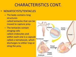
Cnidaria have stinging cells called nematocysts that are used ...
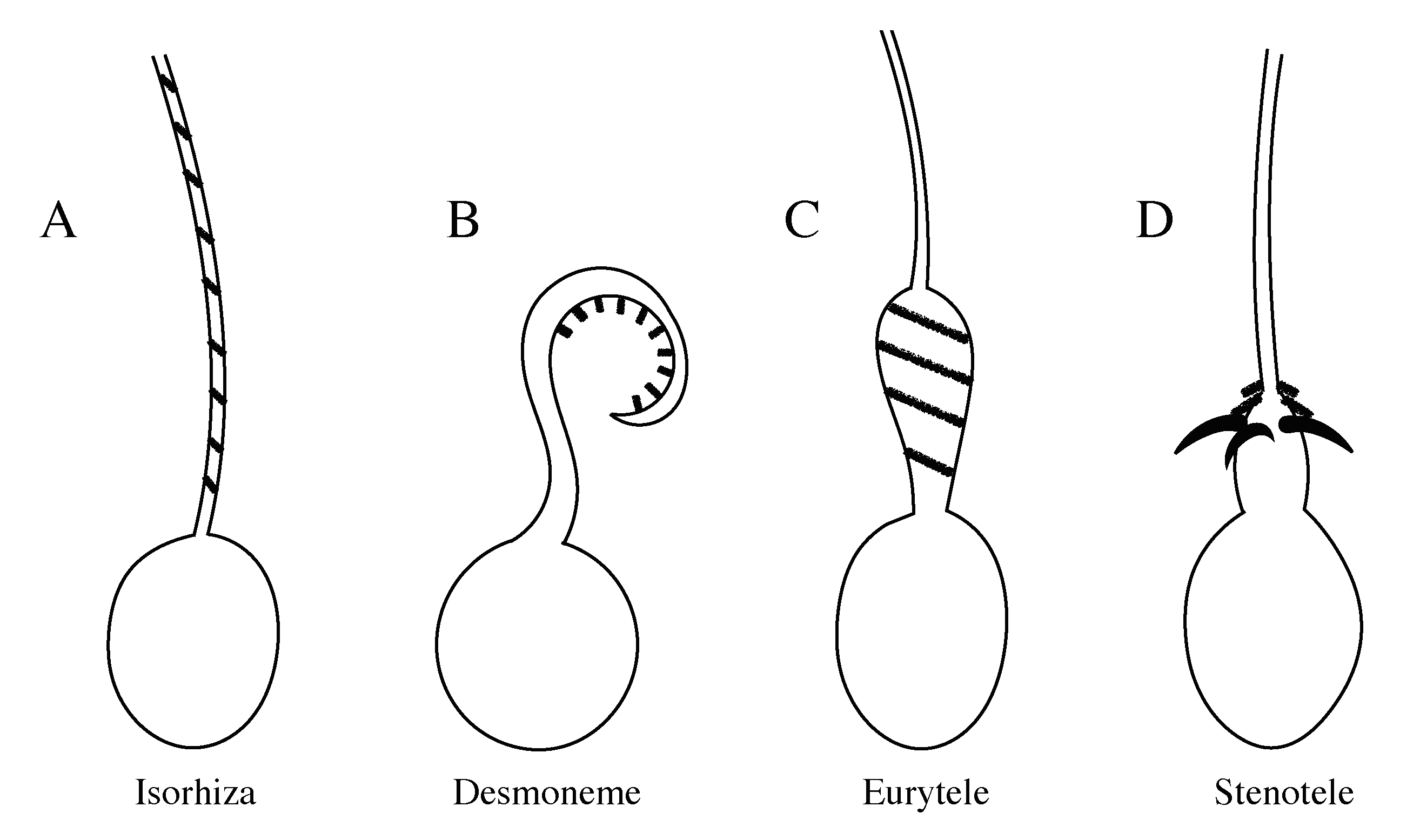
PDF A review of zoanthid nematocyst types and their population structure recognition of nematocyst types, of which at least seven are known for Zoanthidea (Fig. 1), we fol-low Schmidt (1972, 1974), because of his clear diagrams (Fig. 1), but prefer a terminology based on that of Weill (1934). The first group of types comprises: large holotrichs (holotrichs I of Schmidt) or holotrichous isorhizas (i.e. with par-
Cnidocyte - Wikipedia
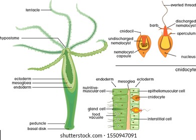
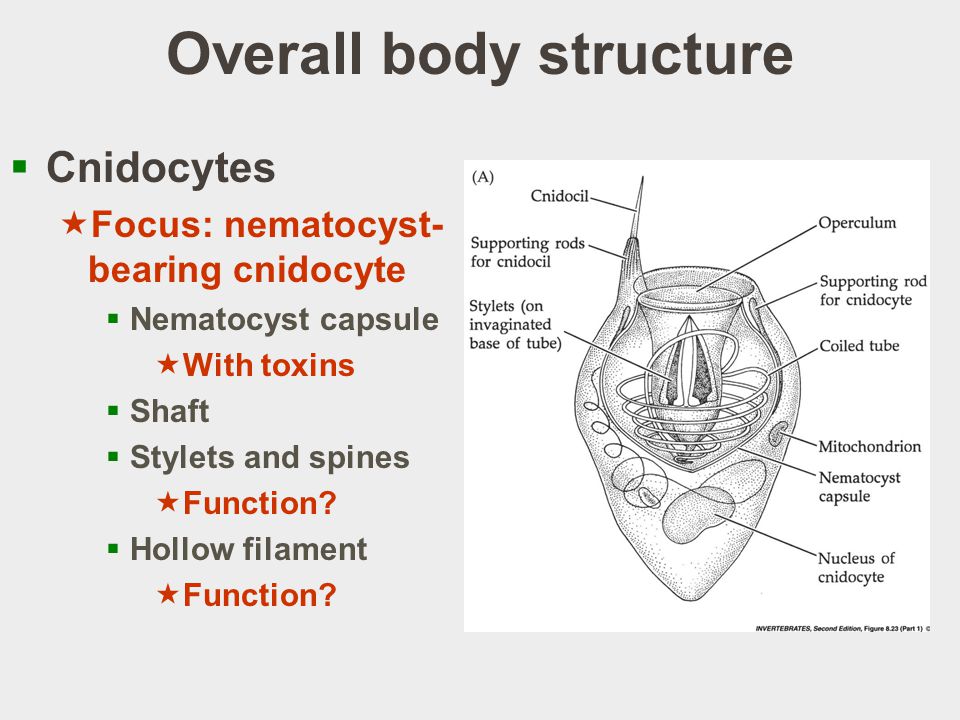
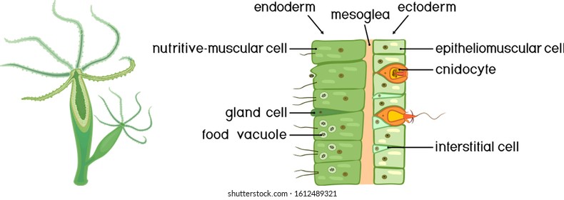
Cnidocyte - Wikipedia A cnidocyte (also known as a cnidoblast or nematocyte) is an explosive cell containing one large secretory organelle called a cnidocyst (also known as a cnida ( pl. cnidae) or nematocyst) that can deliver a sting to other organisms. The presence of this cell defines the phylum Cnidaria ( corals, sea anemones, hydrae, jellyfish, etc.).

Structure of the nematocyst | Download Scientific Diagram
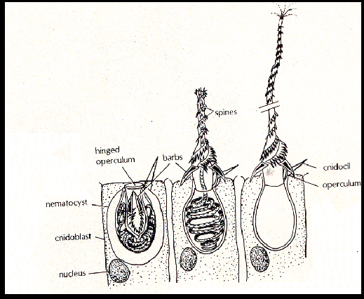
Activity: Nematocysts | manoa.hawaii.edu/ExploringOurFluidEarth Diagram of a cnidocyte ejecting a nematocyst Image by Byron Inouye Fig. 3.28. Anatomy of a sea anemone showing some internal structures. 1. Tentacle, 2. Pharnyx, 5. Septum, 8. Pedal disk, 9. Retractor muscle, 12. Collar, 13. Mouth, 14. Oral disk Image courtesy of Hans Hillewaert, Wikimedia Commons Procedure

What is the function of a lasso in a nematoblast? - Quora
Master Notes on Cnidarians (With Diagram) - Biology Discussion The length of the nematocyst is about 1-12 mm and the length of the discharged tube is about several millimetres long. (ii) The Halistmma colony consists of a long, slender, floating stem and the upper end of the stem bears a small, expanded, gas-filled float or pneumatophore, serves to propel the colony through the water.
Morphological and Molecular Analysis of the Nematostella ...
Functions of Nematocysts in Cnidarians - Study Nature Each individual nematocyst contains a long, hair-like thread with rows of spores at the end. These spore-like structures are built around a coiled tube and have the ability to open pores in prey organisms. The nematocysts vary greatly in shape and they can be either quite small or quite large.
687 Nematocysts Images, Stock Photos & Vectors | Shutterstock
Nematocyst Illustrations & Vectors - Dreamstime Download 24 Nematocyst Stock Illustrations, Vectors & Clipart for FREE or amazingly low rates! New users enjoy 60% OFF. 191,428,014 stock photos online. ... Vector diagram. Structure coral polyp. Coral Anatomy. The coral polyps tend to live in colonies and form the building blocks of the reef. Structure and action cnidocyte. A diagram of the ...
Macrophagy in Hydra | Phylum Cnidaria
Corals Tutorial: Nematocyst Cell - National Ocean Service The diagram above shows the anatomy of a nematocyst cell and its "firing" sequence, from left to right. On the far left is a nematocyst inside its cellular capsule. The cell's thread is coiled under pressure and wrapped around a stinging barb. When potential prey makes contact with the tentacles of a polyp, the nematocyst cell is stimulated.
Biology | Free Full-Text | Quantitative Insights into the ...
Structure of the nematocyst | Download Scientific Diagram - ResearchGate Download scientific diagram | Structure of the nematocyst from publication: Marine and Other Aquatic Dermatoses | Occupational and recreational aquatic activity predisposes our population to a ...
Functional Characteristics of Nematocysts Found on the ...
Nematocyst - Structure, Function, Types and FAQs - VEDANTU Volvent: The volvent, also known as the desmoneme, is indeed a pear-shaped nematocyst. It has a single loop formed by a spineless, short, dense, smooth, and elastic thread tube that is sealed towards the far end. It wraps closely from around prey after being discharged. They seem to be the tiniest nematocysts on the planet.

Volvent Nematocyst, Artwork Wood Print by Francis Leroy ...
Diagrammatic representations of nematocyst types in Palythoa ... Diagrammatic representations of nematocyst types in Palythoa tuberculosa (the same types are found in Protopalythoa), exploded and unexploded. A,A', Large holotrich (A at one-third scale), scapus....

Contribution to the understanding of seasonal cycle of ...
Structure of a nematocyst. cnidocyte. Vector diagram - Shutterstock Structure Nematocyst Cnidocyte Vector Diagram Stock Vector (Royalty Free) 176251196 Download for free See more Popularity score High Usage score High usage Superstar Shutterstock customers love this asset! Item ID: 176251196 Structure of a nematocyst. cnidocyte. Vector diagram Formats EPS 5019 × 3910 pixels • 16.7 × 13 in • DPI 300 • JPG
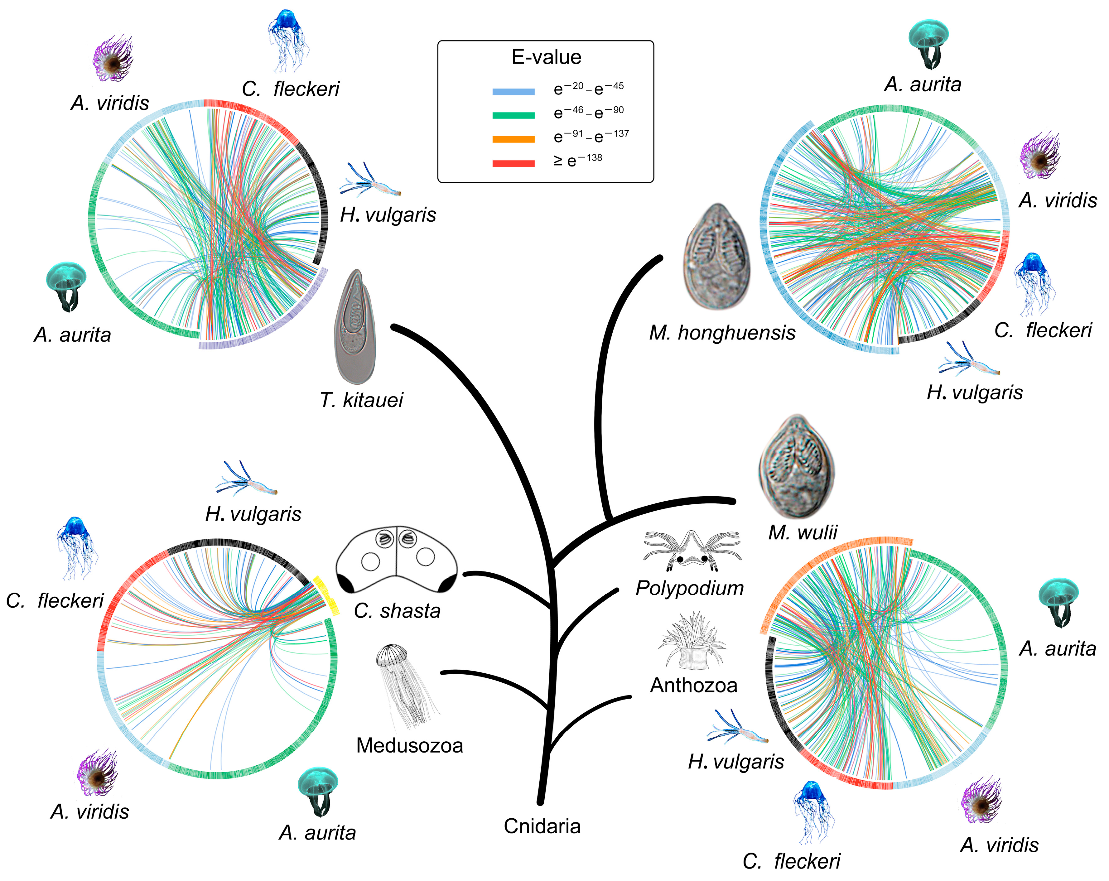
Tree of Life The tree of life according
Nematocyst Definition and Examples - Biology Online Dictionary A nematocyst diagram can be seen in Figure 1 below. Figure 1: Diagram of nematocysts in coral. Image Credit: NOAA.gov. In cnidarians, nematocysts are important in the action of locomotion as well as prey capturing and defense mechanisms. These are seen in jellyfish nematocysts and many others.
Rob's Marine Moment::
Phylum cnidaria
Lecture 6: Phylum Cnidaria, Part 1 - ppt download
Cnidarians
687 Nematocysts Images, Stock Photos & Vectors | Shutterstock

Classification - Science Assessment Term 3
687 Nematocysts Images, Stock Photos & Vectors | Shutterstock
Fluids | Free Full-Text | Fluid Dynamics of Ballistic ...

Nematocyst Definition and Examples - Biology Online Dictionary

Figure 1 from Ultrastructure of the dinoflagellate Polykrikos ...
Prey capture ecology of the Cubozoan Carukia barnesi |

A comparison of the structure and function of nematocysts in ...
Body Wall and Nematocysts - Swaggology

The Silent Sentinels - the Demise of Tropical Coral Reefs

Learn About Nematocyst | Chegg.com
New class of crosslinker-free nanofiber biomaterials from ...

KSLOF Coral Reef Education Portal: Free Coral Feeding ...
Phylum Cnidaria | Biology I | | Course Hero

Phylum Cnidaria /Coelentrata | Characteristics, Classification, Cnidocytes, Nematocyst of Cnidarians

Biology, Biological Diversity, Invertebrates, Phylum Cnidaria ...

Model of elastic nematocyst discharge. (A) schematic ...

nematocyst | biology | Britannica
Corals Tutorial: Nematocyst Cell
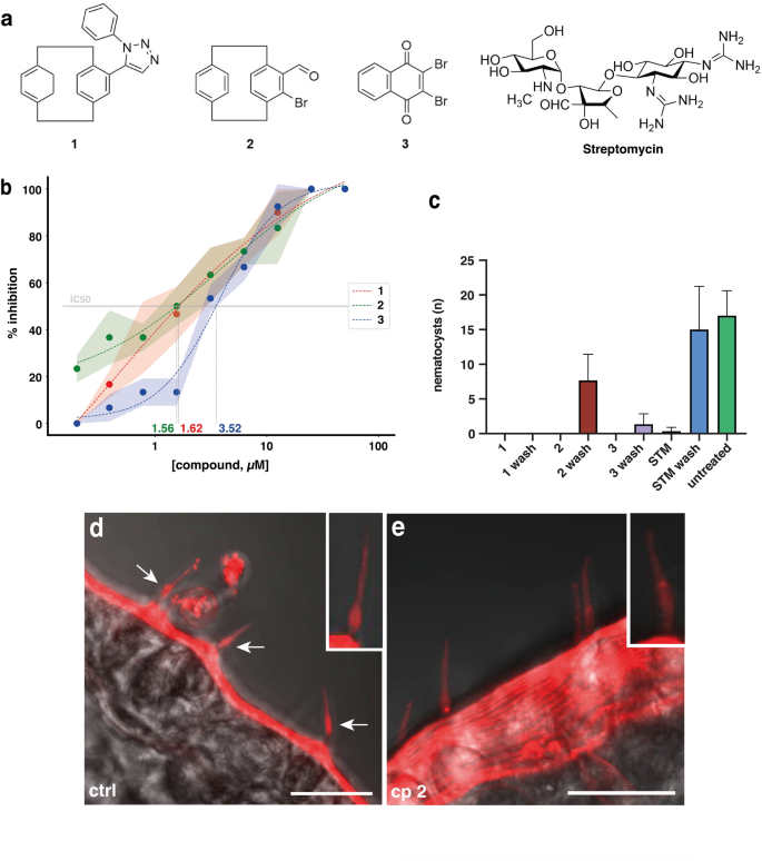
A small molecule screen identifies novel inhibitors of ...
Post a Comment for "38 nematocyst diagram"