38 illuminating parts of microscope
Phase-contrast microscopy - Wikipedia Phase-contrast microscopy (PCM) is an optical microscopy technique that converts phase shifts in light passing through a transparent specimen to brightness changes in the image. Phase shifts themselves are invisible, but become visible when shown as brightness variations. When light waves travel through a medium other than a vacuum, interaction with the medium causes the … Light Microscope vs Electron Microscope: 7 Main Differences Jun 27, 2022 · We’ll break down the different parts of the two microscopes and discuss their features and abilities. Our aim is to illuminate you on all the possibilities the light and the electron microscope bring. Light microscope anatomy. If you’re completely new to the world of microscopes, a light microscope is the best place to start.
PATHOLOGY Synonyms: 17 Synonyms & Antonyms for PATHOLOGY … Find 17 ways to say PATHOLOGY, along with antonyms, related words, and example sentences at Thesaurus.com, the world's most trusted free thesaurus.
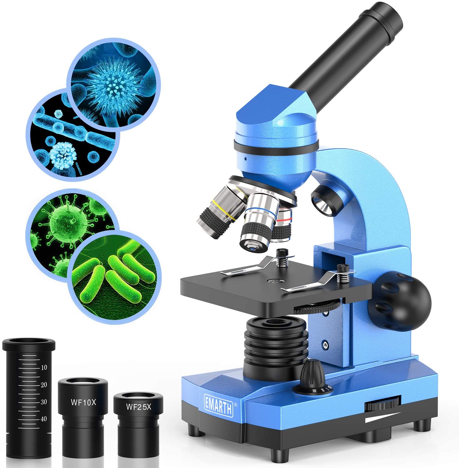
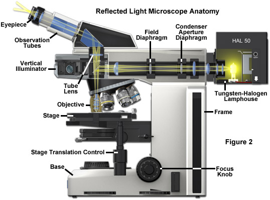
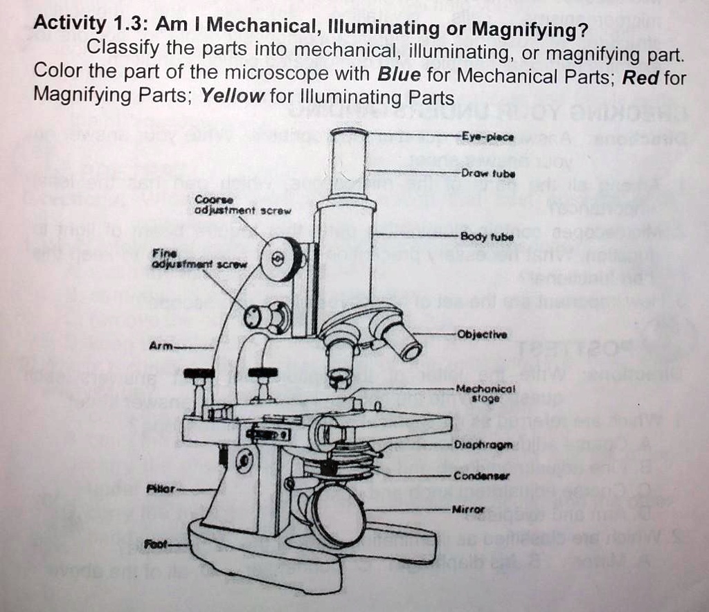
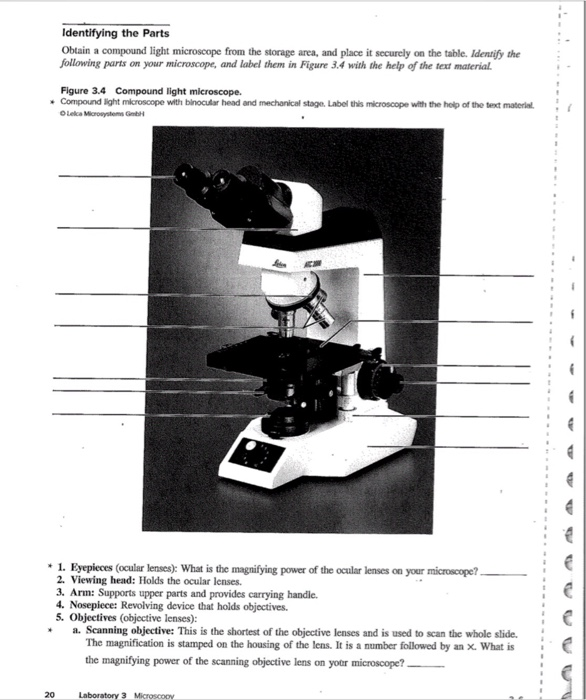
Illuminating parts of microscope
An Introduction to the Light Microscope, Light Microscopy Techniques ... 02/09/2021 · Parts of a microscope and how a light microscope works Simple and compound microscopes Types of light microscopy - Bright field microscopy - Dark field microscopy - Phase contrast microscopy ... Instead of illuminating and imaging the whole sample at once, an illumination spot originating from a point-like source is scanned over the sample area ... Microscope Image Analysis Software | OLYMPUS Stream | Olympus For advanced micro-imaging software that allows you to seamlessly acquire, process, and measure images, our Olympus Stream is the perfect model for you. Electron Microscope: Principle, Types, Applications 19/08/2022 · The major differences between scanning electron microscope (SEM) and transmission electron microscope (TEM) are available here. For Transmission Electron Microscope Specimen Preparation . The specimen suitable for electron microscopes should be very thin (20-100 nm thickness) so the bacterial cells and any other biopsy materials should be …
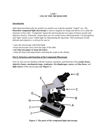
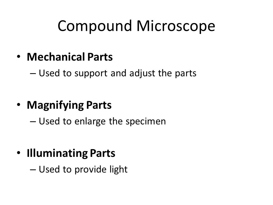
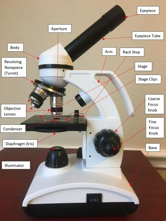
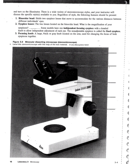
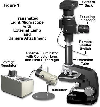
Illuminating parts of microscope. How does a Microscope work The resolution is determined by the frequency of the light waves illuminating the specimen and the quality of the lens. A rule of optical physics is that the shorter the wave length the greater the resolution. Usually expressed in microns, the best resolution a light microscope can produce is 0.2 microns or 200 nanometers. Looking at the Structure of Cells in the Microscope The microscope is generally used with fluorescence optics (see Figure 9-12), but instead of illuminating the whole specimen at once, in the usual way, the optical system at any instant focuses a spot of light onto a single point at a specific depth in the specimen. A very bright source of pinpoint illumination is required; this is usually ... Compound Microscope – Diagram (Parts labelled), Principle and … 03/02/2022 · The three structural components include: 1. Head – This is the upper part of the microscope that houses the optical parts 2. Arm – This part connects the head with the base and provides stability to the microscope. Arm is used to carry the microscope around 3. Base – Base is on which the microscope rests and the base houses the illuminator that lights up the … Confocal microscopy - Wikipedia Confocal microscopy, most frequently confocal laser scanning microscopy (CLSM) or laser confocal scanning microscopy (LCSM), is an optical imaging technique for increasing optical resolution and contrast of a micrograph by means of using a spatial pinhole to block out-of-focus light in image formation. Capturing multiple two-dimensional images at different depths in a …
Electron Microscope: Principle, Types, Applications 19/08/2022 · The major differences between scanning electron microscope (SEM) and transmission electron microscope (TEM) are available here. For Transmission Electron Microscope Specimen Preparation . The specimen suitable for electron microscopes should be very thin (20-100 nm thickness) so the bacterial cells and any other biopsy materials should be … Microscope Image Analysis Software | OLYMPUS Stream | Olympus For advanced micro-imaging software that allows you to seamlessly acquire, process, and measure images, our Olympus Stream is the perfect model for you. An Introduction to the Light Microscope, Light Microscopy Techniques ... 02/09/2021 · Parts of a microscope and how a light microscope works Simple and compound microscopes Types of light microscopy - Bright field microscopy - Dark field microscopy - Phase contrast microscopy ... Instead of illuminating and imaging the whole sample at once, an illumination spot originating from a point-like source is scanned over the sample area ...












Post a Comment for "38 illuminating parts of microscope"