43 microscope pictures with labels
Labeling the Parts of the Microscope Labeling the Parts of the Microscope This activity has been designed for use in homes and schools. Each microscope layout (both blank and the version with answers) are available as PDF downloads. You can view a more in-depth review of each part of the microscope here. Download the Label the Parts of the Microscope PDF printable version here. Microscope, Microscope Parts, Labeled Diagram, and Functions Revolving Nosepiece or Turret: Turret is the part of the microscope that holds two or multiple objective lenses and helps to rotate objective lenses and also helps to easily change power. Objective Lenses: Three are 3 or 4 objective lenses on a microscope. The objective lenses almost always consist of 4x, 10x, 40x and 100x powers. The most common eyepiece lens is 10x and when it coupled with ...
Plant Cell Under Microscope 40X Labeled : 1 - Chloroplast and cell wall ... 1.can only turn fine adjustment 2.draw one row of cells across the middle 3.label the chloroplasts and cell wall. Eukaryotic cells found in viridiplantae; Onion cell) magnification (40x, 100x, or 400x) label all leaf cell under microscope labeled written by macpride monday, april 13, 2020 add. Microscope comes in different types that produce ...

Microscope pictures with labels
Microscope Labeled Pictures, Images and Stock Photos Browse 48 microscope labeled stock photos and images available, or start a new search to explore more stock photos and images. Newest results Fluorescent Imaging immunofluorescence of cancer cells growing... Plant Tissue Systems vector illustration. Labeled biology... Microscope diagram vector illustration. Labeled zoom instrument... Label the microscope — Science Learning Hub In this interactive, you can label the different parts of a microscope. Use this with the Microscope parts activity to help students identify and label the main parts of a microscope and then describe their functions. Drag and drop the text labels onto the microscope diagram. Interactive Bacteria Cell Model - CELLS alive Ribosomes: Ribosomes give the cytoplasm of bacteria a granular appearance in electron micrographs.Though smaller than the ribosomes in eukaryotic cells, these inclusions have a similar function in translating the genetic message in messenger RNA into the production of peptide sequences (proteins).
Microscope pictures with labels. Microscope Labeling Game - PurposeGames.com About this Quiz. This is an online quiz called Microscope Labeling Game. There is a printable worksheet available for download here so you can take the quiz with pen and paper. This quiz has tags. Click on the tags below to find other quizzes on the same subject. Science. Microscope illustrations and clipart (68,272) - Can Stock Photo Microscope Illustrations and Stock Art. 67,723 Microscope illustration and vector EPS clipart graphics available to search from thousands of royalty free stock clip art designers. Content Type All Images Photos Illustrations Vectors Video Specific Orientation People Search With People Without People Exclude From Results Search Type Robert Hooke - Biography, Facts and Pictures Robert Hooke was a Renaissance Man - a jack of all trades, and a master of many. He wrote one of the most significant scientific books ever written, Micrographia, and made contributions to human knowledge spanning Architecture, Astronomy, Biology, Chemistry, Physics, Surveying & Map Making, and the design and construction of scientific instruments. Compound Microscope Parts, Functions, and Labeled Diagram Compound Microscope Definitions for Labels. Eyepiece (ocular lens) with or without Pointer: The part that is looked through at the top of the compound microscope. Eyepieces typically have a magnification between 5x & 30x. Monocular or Binocular Head: Structural support that holds & connects the eyepieces to the objective lenses.
Fluorescence - Wikipedia Fluorescence is the emission of light by a substance that has absorbed light or other electromagnetic radiation.It is a form of luminescence.In most cases, the emitted light has a longer wavelength, and therefore a lower photon energy, than the absorbed radiation. PDF Parts of a Microscope Printables - Homeschool Creations Label the parts of the microscope. You can use the word bank below to fill in the blanks or cut and paste the words at the bottom. Microscope Created by Jolanthe @ HomeschoolCreations.net. Parts of a eyepiece arm stageclips nosepiece focusing knobs illuminator stage objective lenses Nancy Drew 27: The Deadly Device walkthrough - GameBoomers The text looks like video labels. Click on the monitor and then security icon. Enter the text: LBTY6510259. (Thanks, misslilo). See a video of Niko entering the Tesla coil lab and then Gray exiting. After that the blast inside the lab occurred. A binary code appeared at right: 11101001. We need help here. Microscope Images at Various Magnifications | Microscope World Resources The compound microscope typically has three or four magnifications - 40x, 100x, 400x, and sometimes 1000x. At 40x magnification you will be able to see 5mm. At 100x magnification you will be able to see 2mm. At 400x magnification you will be able to see 0.45mm, or 450 microns. At 1000x magnification you will be able to see 0.180mm, or 180 microns.
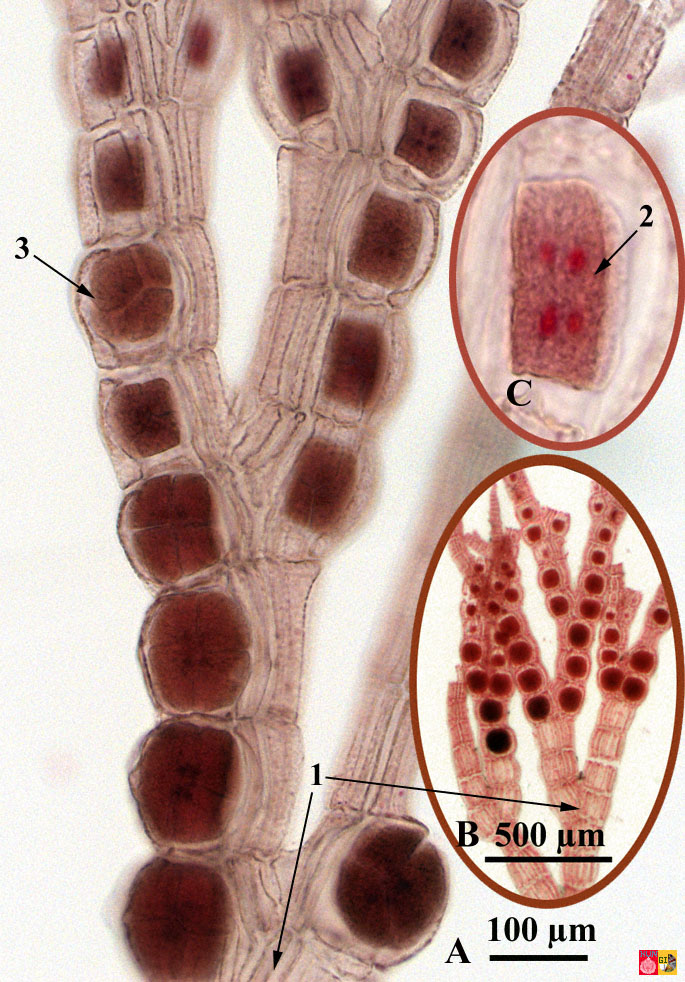
26+ Picture Of A Microscope With Label PNG 26+ Picture Of A Microscope With Label PNG. Microscopes are specially created to magnify the image of the subject being studied. Students label the parts of the microscope in this photo of a basic laboratory light microscope. Microscope Drawing And Label at GetDrawings | Free download from getdrawings.com I searched for this on bing.com/images. Microscope Diagram and Quiz | Science printables, Microscope parts ... Download them all in one convenient PDF, and select the version that's best for your classroom. This PDF contains the following: 1. Parts of a Microscope Diagram - Color 2. Parts of a Microscope Diagram - Black and White 3. Blank Parts of a Microscope Diagram - Black and White 4. Blank, Unlabeled Parts of a Microscope Diagram - Black and White 5. Microscope Images, Stock Photos & Vectors | Shutterstock 457,612 microscope stock photos, vectors, and illustrations are available royalty-free. See microscope stock video clips Image type Orientation Color People Artists More Sort by Jobs/Professions Healthcare and Medical medicine research Turn on AI Powered Search Set goals and get predicted insights based on performance. Close Amazing 27 Things Under The Microscope With Diagrams Amazing 27 Things Under The Microscope With Diagrams February 20, 2022 by Anupama Sapkota Note: Each image source is given below in this post of respective subheadings. Table of Contents 1. Amoeba under the microscope Direct observation Observation after staining 2. Algae under the microscope Chlorophyta Chromophyta Cryptophyta Rhodophyta
ᐈ A labeled microscope stock pictures, Royalty Free ... - Depositphotos ⬇ Download microscope - stock picture and photos in the best photography agency reasonable prices millions of high quality and royalty-free stock photos and images.
Microscope picture label Flashcards | Quizlet Microscope picture label Flashcards | Quizlet Microscope picture label STUDY Flashcards Learn Write Spell Test PLAY Match Gravity Created by kfire Terms in this set (12) Arm What is the part labelled C? Base What is the part labelled D? Body tube What is the part labelled B? Ocular lens What is the part labelled A? Illuminator
Canon U.S.A., Inc. | EOS Utility Download Instructions. Select your model on the left side of this page. Once you arrive at the product support page follow these instructions. Select the “Drivers & Downloads” tab
The Parts of a Microscope (Labeled) Printable - TeacherVision The Parts of a Microscope (Labeled) Printable. Download. Add to Favorites. Share. This diagram labels and explains the function of each part of a microscope. Use this printable as a handout or transparency to help prepare students for working with laboratory equipment. Grade:
Label a Microscope - Storyboard That Create a poster that labels the parts of a microscope and includes descriptions of what each part does. Click "Start Assignment". Use a landscape poster layout (large or small). Search for a diagram of a microscope. Using arrows and textables label each part of the microscope and describe its function. Copy This Storyboard* More options
Pictures of Demodex Mites in Adults and Children - ovante Apr 24, 2017 · First Stage of Demodicosis (Demodex) in Humans shown on pictures (images) belowThe initial or First Stage of Demodicosis in Humans is impossible to see with naked eye. Only your doctor can tell if you have demodex mites on the skin. When you visit a dermatologist, ask him to check the skin scraping under a microscope for positive diagnosis.
Parts of a microscope with functions and labeled diagram Optical parts of a microscope and their functions The optical parts of the microscope are used to view, magnify, and produce an image from a specimen placed on a slide. These parts include: Eyepiece - also known as the ocular. This is the part used to look through the microscope. Its found at the top of the microscope.
Microscope Parts and Functions Microscope Parts and Functions With Labeled Diagram and Functions How does a Compound Microscope Work?. Before exploring microscope parts and functions, you should probably understand that the compound light microscope is more complicated than just a microscope with more than one lens.. First, the purpose of a microscope is to magnify a small object or to magnify the fine details of a larger ...
300+ Free Microscope & Laboratory Images - Pixabay Upload 399 Free images of Microscope Related Images: laboratory science bacteria research scientist lab biology chemistry medical Find your perfect microscope image. Free pictures to download and use in your next project. 399 Free images of Microscope / 4‹ ›


Post a Comment for "43 microscope pictures with labels"